Inoue balloon over left ventricle
Image in Interventional Cardiovascular Medicine: what is displayed on this image? ... and do not hesitate to comment how you would treat this case !
A 48 year-old female patient, diabetic, presented with a history of chest pain over one week.The ECG showed T inversion in leads I, aVL, 2D Echocardiogram showed mild LV systolic dysfunction, RWMA in LCx territory and an 3.4 x 2.8 cms outpouching from inferolateral wall of LV with a narrow neck.
An image is worth a 1,000 words: tell us what you see in this image!
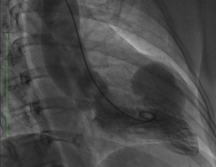
48 year-old female patient, diabetic, presented with history of chest pain over one week.
Authors
Sivalingam Jegan1, Alagarsamy Mathavan1- Hannah Joseph Hospital, Tamil Nadu, INDIA
No comments yet!