A detailed and comprehensive description of the anatomy of the aortic valvar complex as it pertains to TAVI
While there are several publications describing the anatomy of the aortic valvar complex, few specifically focus on the unique challenges presented by transcatheter aortic valve implantation (TAVI). Here you will find a detailed and comprehensive description of the following:
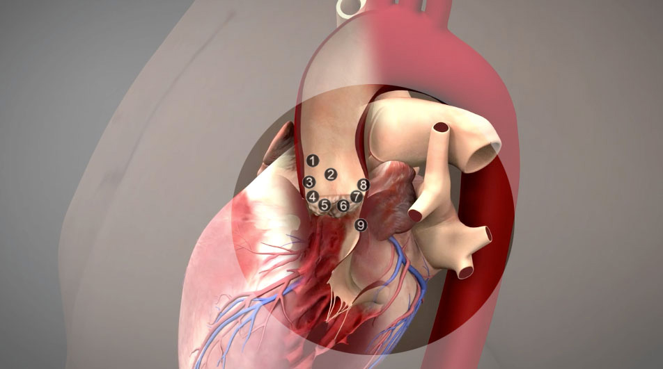
Image courtesy of Edwards Lifesciences LLC, Irvine, CA
- Sinotubular junction: The superior attachments of the aortic valvar leaflets demarcate the level of the sinotubular junction. This junction marks the exit of the aortic root, and the beginning of the ascending aorta.
- Aortic root: The aortic root refers to the aortic outflow tract from its entrance at the left ventricular outlet to its junction with the ascending portion of the aorta.
- Coronary arteries: The coronary arteries arise from the aortic sinuses. The initial portion of the aortic root, which houses the leaflets of the aortic valve, is occupied by the aortic sinuses, also called the sinuses of Valsalva.
- Aortic sinuses: An aortic sinus is one of the anatomic dilations of the ascending aorta, which occurs just above the aortic valve. These widenings are between the wall of the aorta and each of the three cusps of the aortic valve.
- Aortic leaflets: The normal aortic valve is trifoliate, the three leaflets form the aortic valve and provide its main sealing mechanism.
- Bicuspid aortic valve: The aortic valve with two leaflets, usually said to be bicuspid, is the most commonly recognised form of adult congenital heart disease.
- Aortic annulus: The aortic annulus is a fibrous ring at the aortic orifice to the front and right of the atrioventricular aortic valve and is considered the transition point between the left ventricle and aortic root. The annulus is part of the fibrous skeleton of the heart.
- Aortic root diameters: Normalized diameters for the dynamic aortic root vary among the age groups. There are apparent relationships between the dynamic diameters and age, body surface area, weight, and height of the patient.
- Relationship to the conduction system: The conducting system of the heart consists of cardiac muscle cells and conducting fibers (not nervous tissue) that are specialized for initiating impulses and conducting them rapidly through the heart.
This content was originally published as part of The TAVI Atlas, by Darren Mylotte, Diane E. Spicer, Anne E. Sarwark, Carl L. Backer, Robert H. Anderson and Nicolo Piazza