Strange image in the right ventricle wall
Image in interventional cardiovascular medicine: what is displayed on this image? ... And do not hesitate to comment on how you would treat this case!
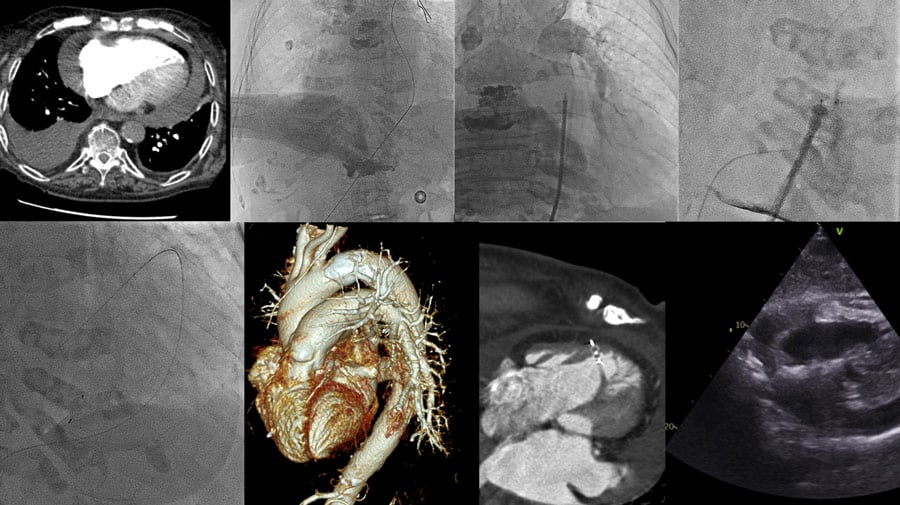
This is a CT scan performed 7 days after the onset of the symptoms. We can see a bilobal image in the right ventricle wall and also in the pericardial space and in the ventricle cavity.
An image is worth a 1,000 words: participate in the quiz below to tell us what you see in this image!
Strange image in the right ventricle wall
Authors
- GHMF, Grenoble, FRANCE
2 comments
Great case. We tried it with Perclose ProGlide™, but not successful. And published it as ............. Vijayvergiya R, Shrimanth YS, Kasinadhuni G, Singh H, Sharma A, Bhargav A, Kaur N. Percutaneous suture based device closure of an inadvertent right ventricle perforation following pericardiocentesis. Anatol J Cardiol. 2021 Nov;25(11):829-831. doi: 10.5152/AnatolJCardiol.2021.49. PMID: 34734817; PMCID: PMC8575405.
Nice case. Using amplatzer device to seal iatrogenic rv puncture during pericardiocentsis. It has been reported quite a few times. We practice keeping 014 wire into the ventricle besides the device just for safety in case device jumps out.