Useful retrieval devices to recover a dislodged or damaged coronary stent
Coronary stent loss complications
The armamentarium of today’s interventionalist is vast and in constant evolution, all the more reason that it is important when dealing with complications to have a quick and comprehensive reference. This section provides just that, allowing you to explore the different tools available for recovering lost or damaged stents and other devices. Here you will find descriptions – and illustrations – of what devices you can use today…
Table of content
Tools available for foreign body retrieval have rapidly evolved in the past decade. Below are a few examples:
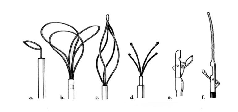
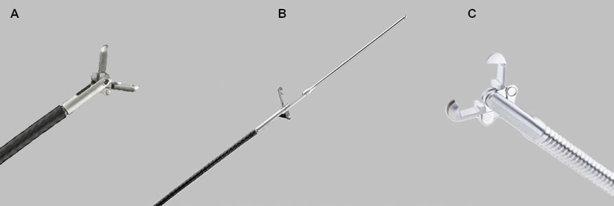
Figure 42.2 Retrieval devices for lost stents: A) Loop snare, B) En-snare three-loop retrieval system, C) Basket retrieval device, D) Biliary stone forceps, E) Biopsy forceps, F) Cook retained-fragment retriever
Loop snares
True 90° snare loop remains coaxial to the lumen:
- Snare loop forms a true 90° angle.
- Device remains coaxial to the lumen for proper insertion and successful retrieval or manipulation of atraumatic foreign bodies.
Snares are available in a range of sizes to ensure optimal fit in the cardiovascular system or hollow viscus, for more information, please consult this comparison table on stent snares and forceps. Proper sizing of the snare prior to procedure ensures straightforward retrieval in target vessel and less manipulation time within the vessel.
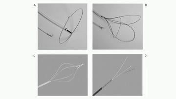
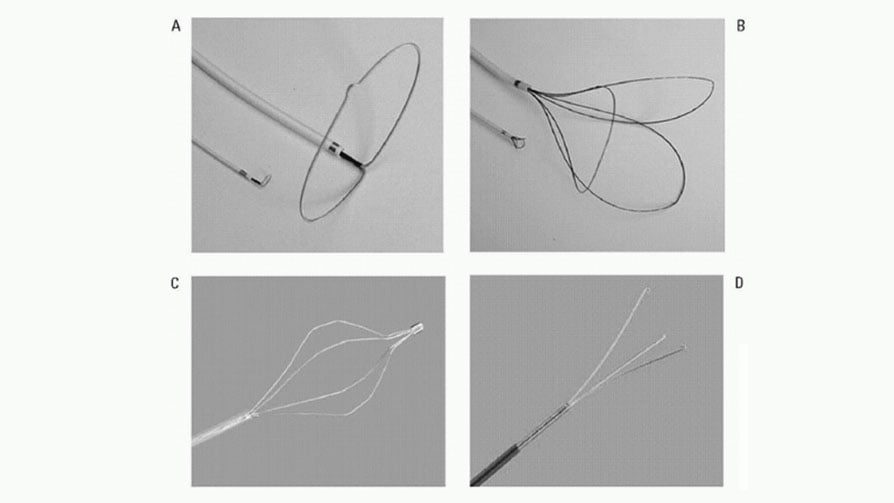
A) Single-loop snare (ONE Snare, Merit Medical)
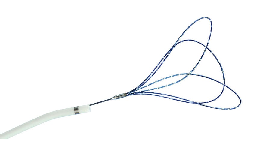
B) Triple-loop snare (EN Snare, Merit Medical)
C) Retrieval basket (Cook Medical Inc.)
D) Grasping Forceps (Cook Medical Inc.)
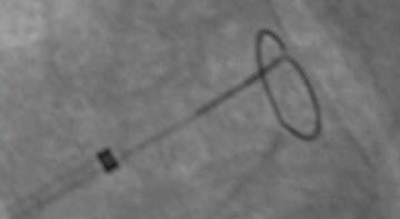
One snare visible under fluoroscopy
The MICRO Elite Snare is intended for use in the retrieval and manipulation of atraumatic foreign bodies located in the coronary and peripheral cardiovascular system and the extra-cranial neurovascular anatomy. This is the only 0.014'' compatible guidewire-based snare.
Micro Elite Snare

Micro Elite Snare: a 0.014" compatible guidewire-based snare
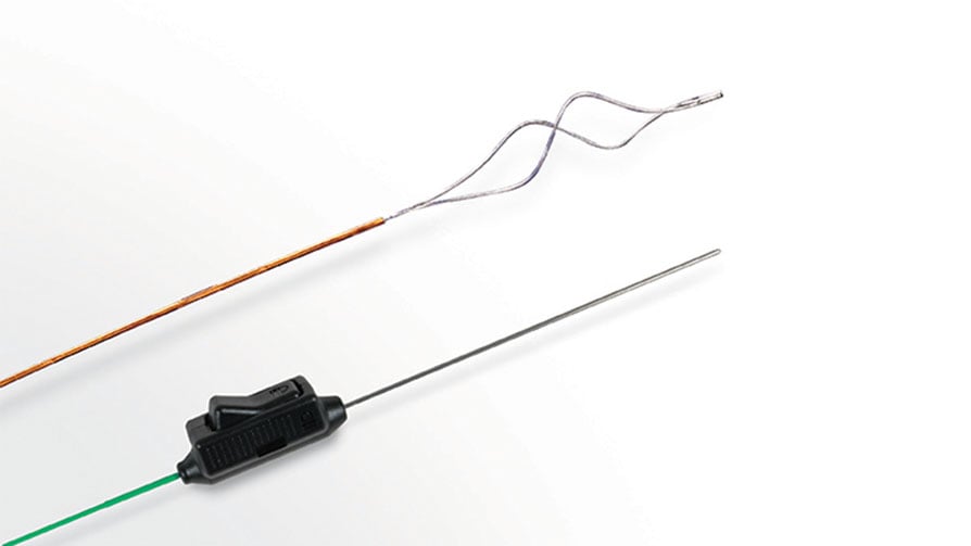
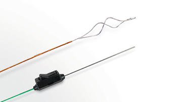
En-snare three-loop retrieval system
The EN Snare Endovascular Snare System is designed with three interlaced loops to retrieve and manipulate foreign objects in the body. The EN Snare can be used to retrieve inferior vena cava filters, reposition indwelling venous catheters, fibrin sheath stripping, or to assist in central venal access venipuncture (Figure 42.2 B). Super elastic Nitinol provides flexibility, kink resistance, and torque control. The EN Snare is designed to rotate and expand for excellent coverage and retrieval within a variety of vessel sizes. Platinum strands are incorporated into the EN Snare loops for excellent visualization under fluoroscopy.
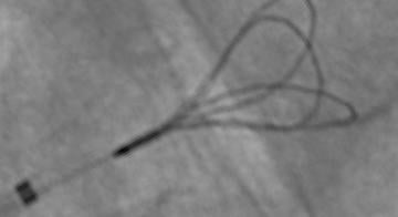
EN Snare endovascular snare system visible under fluoroscopy

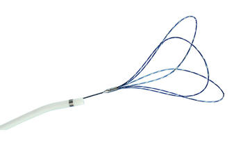
EN Snare Endovascular Snare System
For more information about snares, please consult:
- Medtronic - Microsnare
- Merit Medical - EN Snare® Endovascular Snare System
- Vascular Solutions - MICRO Elite™ Snare
Basket retrieval devices
The basket retrieval device (Figure 42.2 C) consists of a hellically arrange loops that can be expanded or collapsed. Originally designed to remove billiary stones, this device could also be used to trap lost or retained components within its loops, for instance catching a stent.
Forceps
Retrieval using forceps are feasible if the stent is present within the most proximal part of the left main or right coronary arteries but cannot be recommended as a first line option as it is aggressive. If employed then use of small French (paediatric sized) myocardial bioptomes is best (3Fr-1.3mm). Alternatively a biliary forceps can be employed. Forceps can also be used from the homolateral or contralateral femoral sheath once the retained material is brought down to the level of the iliac artery. Toothed forceps such as Maslanka grasping forceps may be used.
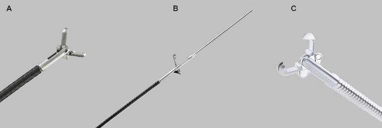
A) Flexible Myocardial Biopsy Forceps (Bioptome), B) Vascular Retrieval Forceps, C) Maslanka Grasping Forceps
For more information about forceps, please consult:
- Cook Medical - Flexible Myocardial Biopsy Forceps (Bioptome)
- Cook Medical - Vascular Retrieval Forceps
- BVM Medical - Maslanka Grasping Forceps
Billiary Stone Forceps
A biliary stone forceps device consists of a set of curved, fingerlike projections that can be expanded/extended or contracted/retracted (Figure 42.2 D). This device was originally designed for the percutaneous removal of stones from the biliary tree. Manipulating the “fingers” of this device, an operator can snag lost or retained components. This device is most useful in recovering partially expanded stents, or in situations in which a portion of a stent has become separated from the deployment balloon catheter. Under these circumstances, it is often possible to remove a damaged stent without having to lose the guidewire position. Biliary forceps are available with catheter bodies of 4 to 5 Fr, and in lengths of 130 cm. The retracted device has fair visibility under fluoroscopy, but the extended fingerlike projections have poor radio-opacity. The device should be used with great caution in the vascular system because laceration of the vessel wall can occur if the retrieved item becomes entrapped in the fingerlike projections of the device.
Myocardial Biopsy Forceps
Another technique involves grasping the material by a myocardial biopsy forceps. If aligned favourably the stent can be removed through the sheath. More often a snare is needed via a contralateral femoral artery.
These devices have distal tip modifications that allow grasping of structures through a “biting jaws” action (Figure 42.2 E). Myocardial biotomes are the most familiar example of such a tool. A variety of forceps are available, but most are not available for vascular use because the shaft diameter is very large, the device is too rigid to be passed safely in the arterial system, and/or the catheter length (often 80 to 90 cm) may be inadequate. The thinner, softer, and longer disposable biotomes may be useful in stent retrieval. Although biotome jaws are not meant for cutting through metal, it is possible that aggressive gripping with such a device could sever a thin metal stent.
Cook Retained-Fragment Retriever
The Cook retained-fragment retriever catheter, manufactured by Cook, Inc. has a guidewire attached to the distal end, resembling a fixed-wire angioplasty balloon catheter (Figure 42.2 F). An articulating arm operable from the proximal hub permits grasping and retrieving of retained equipment fragments. This catheter was developed for use by vascular radiologists and is available in 80 cm and 145 cm lengths.
The Cook retained-fragment retriever is used for retrieval of fragments of catheter tubing, wire guides, and other foreign objects from the vascular system.
This gallery of retrieval devices also contains a number of examples.