Waterball in the heart! - Image 11
Image in Interventional Cardiovascular Medicine: what is displayed on this image? ... and do not hesitate to comment how you would treat this case!
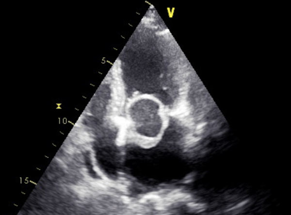
A 49-year-old woman presented with progressive dyspnea for six months. The transthoracic 2D-echocardiogram showed a pedunculated, cystic mass in the left atrium attached to the inter-atrial septum. It measured 3 X 3.3 cm and prolapsed partially through the mitral valve into the left ventricle during diastole causing severe obstruction in transmitral inflow with a mean gradient of 22 mmHg. Due to interatrial septum attachment, possibility of atrial myxoma was considered, but it is unlikely for myxoma to be cystic...
An image is worth a 1000 words: tell us what you see in this image!
Waterball in the heart!
Authors
1 comment
DDs would include - Hydatid cyst, blood cyst, thrombus with central liquifaction