Progressive breathlessness and intracardiac mass
Image in interventional cardiovascular medicine: what is displayed on this image? ... And do not hesitate to comment on how you would treat this case!
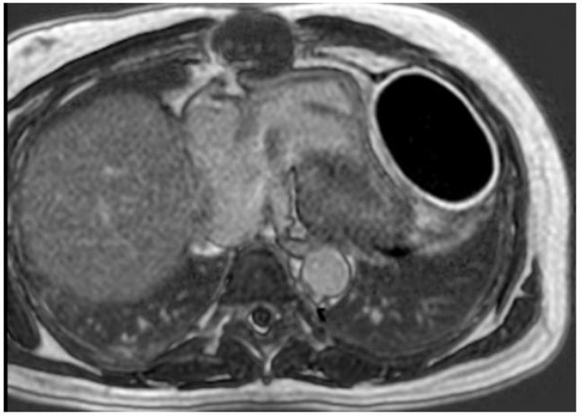
A 65-year-old male patient presented with progressive shortness of breath on exertion for 3 months.He underwent aortic valve replacement using a bioprosthetic valve in another city hospital 6 months ago.Clinical examination revealed pulse rate of 96/min, blood pressure of 110/78 mm Hg, and short systolic ejection murmur on CVS examination.Chest X-ray showed linear calcific opacity on the cardiac silhouette.Echocardiogram showed concentric LV-hypertrophy with good LV function and normal prosthetic valve function with a pericardial mass.Postcontrast cardiac MRI study showed an oval-shaped hypotensive focus in the pericardial region with peripheral edge enhancement (image).
An image is worth a 1,000 words: participate in the quiz below to tell us what you see in this image!

A 65-year-old male patient presented with progressive shortness of breath on exertion for 3 months.
Authors
Baruah Dibyakumar1, Baruah Dibya1, Varma Ravikumar1- Apollo Hospitals, Health City., Visakhapatnam, India


No comments yet!