A different device in the inferior vena cava: what happened?
Image in interventional cardiovascular medicine: what is displayed on this image? ... And do not hesitate to comment on how you would treat this case!
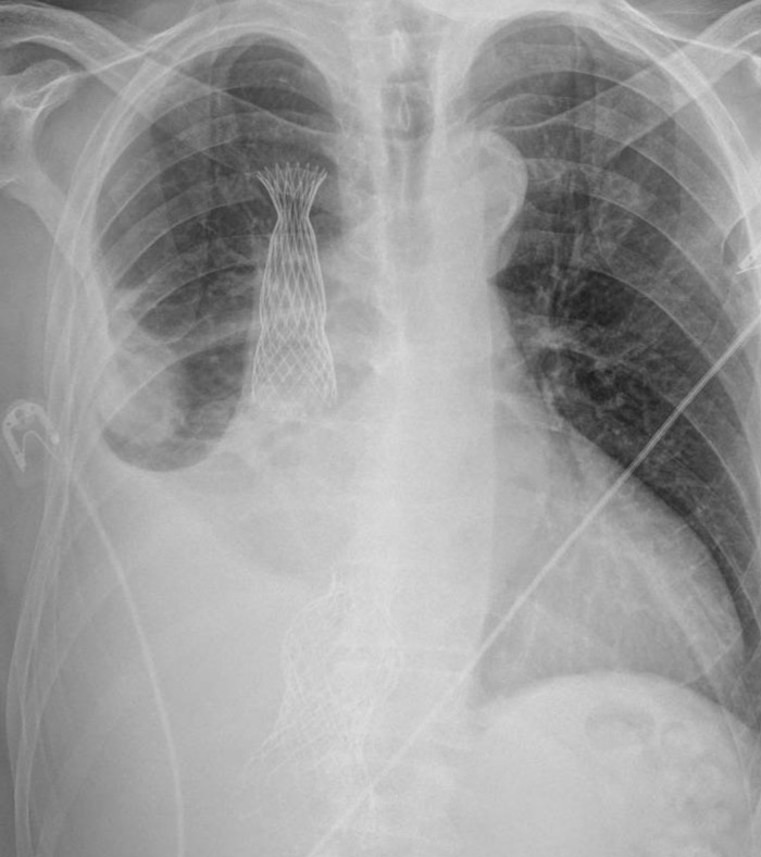
70-year-old man. Heart failure refractory to diuretic therapy. Echocardiogram: severe tricuspid regurgitation with dilated right ventricle. Definition of a bicaval valve implant. During superior vena cava valve implantation, device embolizes right atrium.
An image is worth a 1,000 words: participate in the quiz below to tell us what you see in this image!
A different device in the inferior vena cava: what happened?
Authors
CATALDO Pabla1, DAUVERGNE Christian1, DUARTE Manuel1, CUEVAS Oscar1, MENDEZ Manuel1, PINEDA Fernando1, URIARTE Polentzi1, HERRERA Cecilia1, ARAYA Mario1, SANDOVAL Jorge1
- Hospital del Tórax, Providencia, CHILE
1 comment
Interesting Case. Clearly the first SVC valve was implanted at a low position and embolized. The 2 SVC valve looks at the correct level, that's why did not embolized as the first one. I see the embolized SVC valved is fixed inside the IVC valve. My concern is that this may impair leaflets coaptation of IVC, and the leaflets of the embolized SVC are at the opposite direction of vein flow towards the RA. Maybe a good solution would be to fixate the embolized SVC crown with the second SVC nitinol frame.