Footprints in the aorta
Image in interventional cardiovascular medicine: what is displayed on this image? ... And do not hesitate to comment on how you would treat this case!
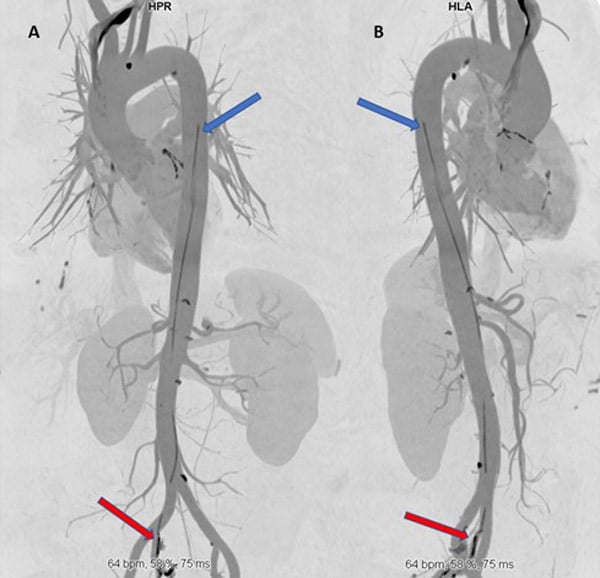
We present the case of a 66-year-old male patient who presented to tertiary medical center emergency department (ED) with abdominal pain. His medical history included ischemic heart disease (IHD) with coronary angiography in 2012, hyperlipidemia, and hypertension. Abdominal ultrasound (US) raised concern of aortic dissection. Computed Tomography (CT) angiography was performed.
An image is worth a 1,000 words: tell us what you see in this image!
Footprints in the aorta
Author
Asher Elad1- Shaare Zedek Medical Center, Jerusalem, ISRAEL
No comments yet!