Patient with dizziness and exertional chest discomfort who ended up with PCI
Image in interventional cardiovascular medicine: what is displayed on this image? ... And do not hesitate to comment on how you would treat this case!
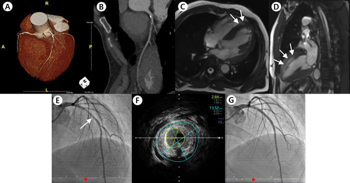
A 58-year-old male patient was referred to the emergency department with a chief complaint of dizziness. Initial vital signs showed a blood pressure of 109/62 mmHg and a heart rate of 44 beats per minute. Brain CT and MRI showed multifocal acute infarctions, but no hemorrhagic findings. Consequently, he was diagnosed with an acute cortex infarction. Besides, the patient explained that he has suffered from severe neuropathic pain since the age of 10. He also complained of post-exercise chest discomfort. Cardiac marker evaluation showed an increased troponin-I level of 219.44 pg/mL, and TTE showed LV hypertrophy with normal ejection fraction. Cardiac MRI using the late gadolinium enhancement technique presented diffuse myocardial fibrosis. With suspicion of Fabry's disease (FD), genetic tests were conducted. As soon as FD was confirmed, we initiated enzyme replacement therapy (ERT). After three months of ERT, PCI was performed since > 70 % stenosis was observed by intravascular ultrasound.An image is worth a 1,000 words: participate in the quiz below to tell us what you see in this image!
Patient with dizziness and exertional chest discomfort who ended up with PCI
Authors
KIM You Jin2, PARK Kyu Tae1, CHOI Hyun Hee1, PARK Soobin2- Hallym University Chuncheon Sacred Heart Hospital, Gangwon-do, REPUBLIC OF KOREA
- Hallym University, Gangwon-do, REPUBLIC OF KOREA
No comments yet!