What's the "Bright Spot" on OCT?
Image in interventional cardiovascular medicine: what is displayed on this image? ... And do not hesitate to comment on how you would treat this case!
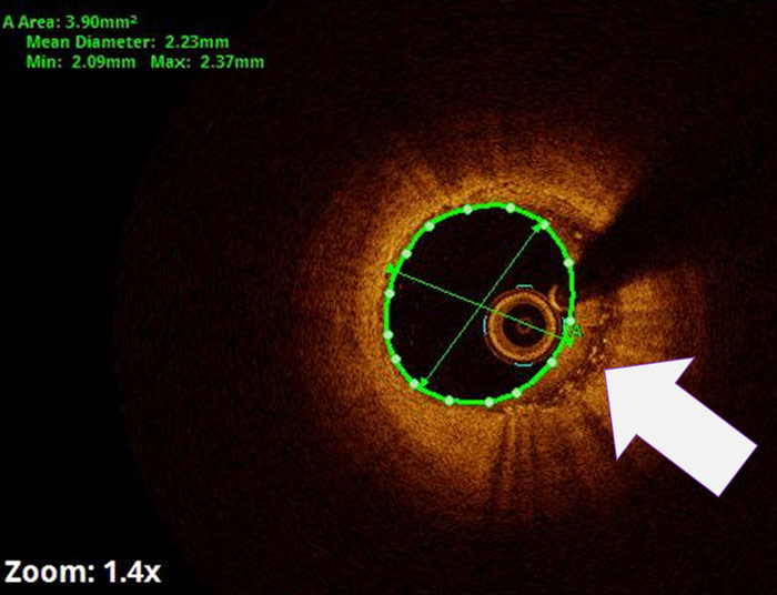
A middle-aged male presented with ACS and intermediate stenosis in LAD. OCT revealed an eccentric plaque with bright spots & MLA of 3.9 mm2. The inflammatory component of the plaque has intrigued physicians about the vulnerability.
An image is worth a 1,000 words: participate in the quiz below to tell us what you see in this image!
What's the "Bright Spot" on OCT?
Authors
BHATIA Tanuj1
- SGRR MEDICAL AND HEALTH sciences college, Uttarakhand, INDIA
3 comments
What do the M1 macrophages look like?
Nice image. Macrophages are not uncommonly found in ACS, if we look carefully.
M1 Macrophages or "Classic" are pro-inflammatory and engulf lipids to form "foam cells", hence appear dark because of a large amount of intracellular lipid. M2 macrophages, on the other hand, are anti-inflammatory having less lipid and more light-scattering mitochondria, hence appearing as "bright".