Focus on Imaging @PCR London Valves 2018
Keep your eyes on what not to miss!
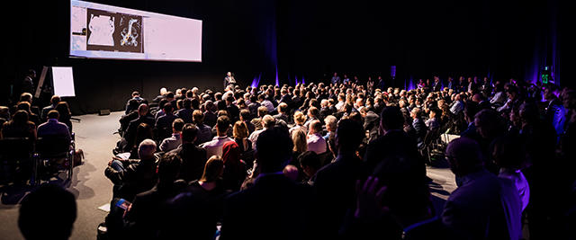
Imaging plays an absolutely essential role in the assessment and treatment of patients with complex heart valve disease. As such PCR London Valves places a strong focus on it throughout the Course, addressing the Heart Team at large: interventional cardiologists, cardiac surgeons, nurses, anaesthesiologists and imaging specialists. The overall Programme notably seeks to teach which skills and knowledge are required to become an interventional imaging specialist, and how to interpret 3D cardiac anatomy using multimodality imaging.
Imaging at a glance
- Kick-off at 11:15 on Sunday with a dedicated Imaging Day for all of the Heart Team
- tutorial sessions
- practical Learning sessions
- case-based Learning sessions
- submission-based sessions (abstracts, clinical cases, e-Posters)
- Wide choice of hands-on imaging sessions in the Training Village, including specific workshops from imaging companies on Sunday
- Every LIVE demonstration on Monday and Tuesday is launched following a 5-minute imaging analysis by a specialist in the techniques used for the case concerned
- NEW this year! Imaging Learning Centre on Monday and Tuesday
Imaging Learning Centre
Join the new structural imaging workshops - “The eyes of the Heart Team”!
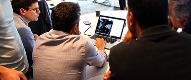
PCR London Valves is delighted to act as forerunner as it hosts the first innovative and practical workshops of this kind, prior to their implementation in other PCR initiatives. Initiated by Course Director and imaging expert Nicolo Piazza, they deal with the correlation between imaging modalities, by demonstrating the use of multi-slice computed tomography as a central tool to better understand fluoroscope and echocardiographic imaging.
Read the article about this new initiative written by Nicolo Piazza.
Key details
- 4 modular structural imaging workshops, allowing participants to select specific ones or attend all four (according to availability and on a first-come first-served basis)
- one workshop introducing heart chamber views and S curves
- 3 subsequent workshops dealing respectively with CT and fluoroscopic anatomy of the aortic, mitral and tricuspid valves
- 90 minutes duration each
- 20 participants max. per workshop