Multimodality imaging in cardiology unveils the rarest diagnostic possibilities!
Image in interventional cardiovascular medicine: what is displayed on this image? ... And do not hesitate to comment on how you would treat this case!
A 26-year-old male patient presented with dry cough, orthopnea, and PND & right-sided heart failure symptoms for 2 weeks. On examination, he has continuous murmur.
2D Echo shows fluid-filled cystic lesion with turbulent flow from aorta & LVEF 33 %
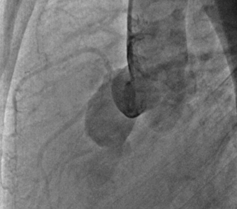
Cardiac MRI & aortic angiogram confirms diagnosis.
An image is worth a 1,000 words: participate in the quiz below to tell us what you see in this image!
Multimodality imaging in cardiology unveils the rarest diagnostic possibilities!
Authors
Mohammad Mobeen Quadri1, Devi Durga1
- Chettinad Hospital And Research Institute, Tamil Nadu, INDIA
No comments yet!