Nightmare in the daylight
Image in interventional cardiovascular medicine: what is displayed on this image? ... And do not hesitate to comment on how you would treat this case!
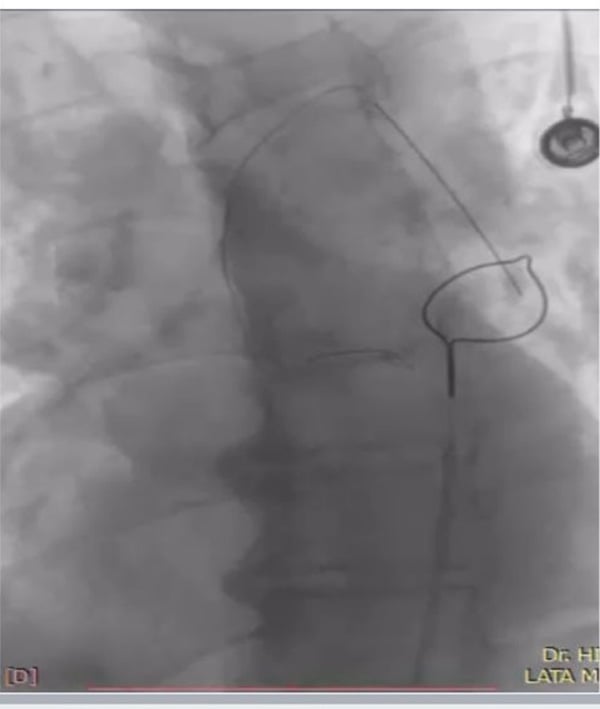
A 62-year-old hypertensive male presented with a 2-day history of chest pain, palpitations, and dyspnea. ECG demonstrated right ventricular hypertrophy, right bundle branch block, and anterior wall ischemia. Echocardiography revealed severe left ventricular dysfunction (EF 20%) with regional wall motion abnormalities in the LAD territory. Coronary angiography identified a heavily calcified chronic total occlusion of the LAD.
A GAIA II wire was used to attempt lesion crossing, but the proximal end became lodged in the ostio-proximal LAD, and the distal tip fractured inside the guide catheter. A balloon catheter was advanced over the wire to retrieve it, but its proximal shaft broke and became stuck within the catheter. During withdrawal, the entire wire configuration was dislodged and suspended in the ascending aorta.
An image is worth a 1,000 words: participate in the quiz below to tell us what you see in this image!
Nightmare in the daylight
Authors
Solanki Shivani1, Bhagwatkar Hitendra1
- NKP Salve Institute of Medical Sciences, Maharashtra, INDIA
1 comment
Single lumen snare