Black and white: tiger stripe!
Image in interventional cardiovascular medicine: what is displayed on this image? ... And do not hesitate to comment on how you would treat this case!
A 70-year-old male presented to the OPD (Outdoor Patient Department) for ECHO (Echocardiography). Patient had a history of breathlessness on walking since past 2-3 months, which would get relieved at rest. He did not have any history suggestive of pedal edema, syncope, chest pain, or palpitations. The patient had history of hypertension since past 3 years, was on irregular medications, and had no other co-morbidities like diabetes mellitus, coronary artery disease.
On auscultation in sitting position, patient had a decrescendo early diastolic blowing murmur heard along the left side of sternum in 3rd intercostal space, which was best audible in leaning forward position in full expiration.
ECG showed LVH with left atrial enlargement and prominent Q waves in l, avL ,V3-V6.
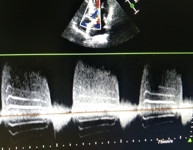
ECHO showed tricuspid aortic valve with mild aortic regurgitation. There was no obvious vegetation or nodules seen on the valves. The pressure half time was 540msec. There was concentric LVH with apical LV aneurysm. The Left ventricle systolic function was normal with EF (Ejection fraction) of 52%. On Color Doppler imaging, there were band like signals seen.
An image is worth a 1,000 words: participate in the quiz below to tell us what you see in this image!
Black and white: tiger stripe!
Author
Bali R.1
- Hero DMC Heart Institute, Ludhiana, Punjab, INDIA
No comments yet!