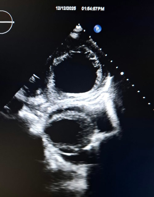
Parasternal short-axis view demonstrating two pulsatile cavities
Image in interventional cardiovascular medicine: what is displayed on this image? ... And do not hesitate to comment on how you would treat this case!
A 71-year-old male with a known history of chronic obstructive airway disease and poor adherence to inhaler therapy presented with a gradual worsening of dyspnea.
A chest X-ray demonstrated hyperinflated lung fields, widening of the mediastinum, and cardiomegaly. His electrocardiogram (ECG) showed sinus rhythm with poor R-wave progression and pathological Q waves in the inferior leads.
Transthoracic echocardiography revealed severe ischemic left ventricular dysfunction, and two pulsatile cavities were visualized in the parasternal short-axis view.
An image is worth a 1,000 words: participate in the quiz below to tell us what you see in this image!
Parasternal short-axis view demonstrating two pulsatile cavities
Authors
Madushanka G.1, Wijethunge N.1, Karunarathne C.1, Premarathne S.1- National Hospital Galle, Galle, SRI LANKA
No comments yet!