Will the real L.A.D., please stand up!
Image in interventional cardiovascular medicine: what is displayed on this image? ... And do not hesitate to comment on how you would treat this case!
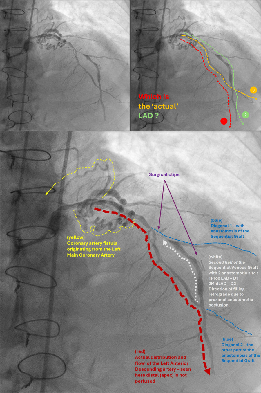
A 70-year-old man underwent coronary angiography for evaluation of new onset chest pain, 15 years after his last major procedure. The first angiography (AP cranial view) after engaging the left main coronary artery, left us stumped.
An image is worth a 1,000 words: participate in the quiz below to tell us what you see in this image!
Will the real L.A.D., please stand up!
Authors
Ramanathan S.1, Bin Zenal Abidin M. A. G.1, Abdullah Ramaiah A. R.1, Selvaraj K.1
- Hospital Serdang, Selangor, MALAYSIA
4 comments
Not able to understand the explanation given in the last image. Can you be little more clear in the explanation? Thank you
Hi all - we submitted this. Kindly focus on the 1st and 2nd image - to determine which is the true LAD Disclaimer : answer is below the 3rd image is the explanation for the answer : 1 TRUE LAD as mentioned 2 Diagnoal 1 and 2 is marked 3 the extra 'LAD' like vessel is actually a sequential Venous ByPass Graft due to occlusion as the first proximal site (pLAD/D1) - it has filled up retrogradely from distal site (mLAD/D2) - mimicking LAD 4 COronary fistula to pulmonary artery 5 distal LAD is still occluded with poor flow I have requested only 1st and 2nd image to be included The 3rd is not serving its purpose.
Hi . We had submitted the case
1 TRUE LAD is as mentioned in the answer 2 Sequential venous bypass graft connecting pLAD/D1 to mLAD/D2 - mimicks another LAD because the pLAD/D1 is occluded , there is retrograde flow from mLAD/D2 upwards 3 dLAD is also occluded , hence no distal flow 4 Coronary fistula present to pulmonary circulation