22 Sep 2023
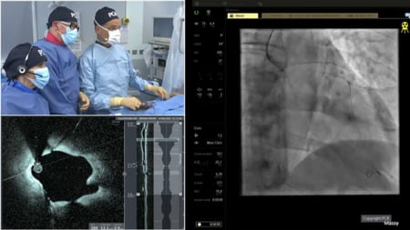
Calcified bifurcation lesion - LIVE case
Provisional stenting with Rotablator, IVL and IVUS-guided drug-eluting balloon
Summary
A 59-year-old female patient with type 2 DM, dyslipidaemia, hypothyroidism and impaired LV function (39%) presented with multivessel disease. RCA PCI was performed 3 days ago.
The prox and mid LAD showed a severe calcified lesion including the diagonal. The lesion was prepared using a 1.25 Rotaglide™ ('single-person' technique), followed by a 2.5 mm IVL.
The bifurcation was treated with an IVUS-guided stent (2.5 - 38 mm). This provisional stenting was completed by DEB kissing in the diagonal ostium and post-dilatation of the proximal LAD.
LIVE case from Apollo Hospital - Chennai, India
Navigate the video by moving your mouse over the chapter icon in the toolbar
Key moments:
- 06:30-08:40 - Angiography analysis
- 18:07-19:52 - Procedural strategy
- 21:00-22:53 - IVUS guidance before Rotablator
- 24:36-29:00 - Rotablator with a 'single-person' technique
- 32:00-34:52 - IVUS guidance after Rotablator
- 49:20-53:58 - IVUS guidance post IVL
- 1:18:30-1:20:30 - Final IVUS
Keywords: Provisional stenting, calcified non-left main bifurcation, Rotablator, IVL, drug-eluting balloon
Learning Objectives
- To understand how intravascular imaging is guiding PCI strategy for treating calcified lesions
- To learn how to use rotational atherectomy and other calcium modification devices for treating calcified lesion
- To define an algorithm for PCI of severely calcified lesion for optimal and safe outcomes

/slide001.jpg)
/slide002.jpg)
/slide003.jpg)
/slide004.jpg)
/slide005.jpg)
/slide006.jpg)
/slide001.jpg)
/slide002.jpg)
/slide003.jpg)
/slide004.jpg)
/slide005.jpg)
/slide006.jpg)
/slide007.jpg)
/slide008.jpg)
/slide009.jpg)
/slide010.jpg)