14 May 2024
How to best image the atrioventricular valves?
With the collaboration of the PCR Tricuspid Focus Group
Anchorperson:
N. Wunderlich
Spokesperson:
A. Latib
Summary
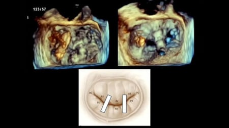
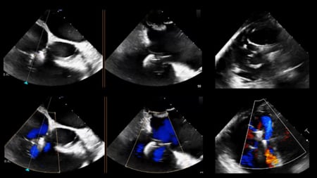
This session, in collaboration with the PCR Tricuspid Focus Group, aims to provide a comprehensive understanding of how to image the atrioventricular valves (mitral and tricuspid) to assess their anatomy and pathology. It covers the step-by-step evaluation of the mitral and tricuspid valves using transesophageal echocardiography (TEE) and the additional information that can be obtained from CT scans. The session also discusses the most important imaging information required for the different transcatheter treatment strategies for the mitral and tricuspid valves, emphasizing the crucial role of imaging in patient selection and procedural planning.
Learning Objectives
- To learn how to image the atrioventricular valves to understand the anatomy and pathology by echo
- To learn what imaging information is needed for optimal patient selection for atrioventricular valves procedures
- To learn what information CT can contribute to patient selection for atrioventricular valves procedures