24 Feb 2020
Ultrasound-guided puncture of the femoral artery for TAVI
Ultrasound can enhance safety and facilitate femoral puncture... Discover the tips and solutions proposed by Won-Keun Kim!
Frequency of the problem:
Expert level:
Summary
Ultrasound can enhance safety and facilitate femoral puncture.
The problem
To obtain safe femoral access for the insertion of large-bore sheaths, there are a few points that should be observed:
- Target the common femoral artery above the femoral bifurcation, the needle should enter the vessel in an orthogonal angle to the cross-section, otherwise closure devices may not work appropriately!
- Puncture site should be in projection to the femoral head (Figure 1), otherwise manual compression may be less effective.
- Puncture too high → risk of retroperitoneal bleeding.
- Puncture too low → risk of pseudoaneurysm!
Fluoroscopy – either with or without angiographic overlay - is the most commonly used approach for femoral puncture, but is associated with radiation exposure and may be less suitable to achieve an orthogonal puncture angle (Video 1).
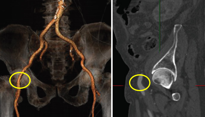
Figure 1.
Principal idea
Ultrasound is a safe and ubiquitously available imaging modality which provides a detailed visualization of the target anatomy to guide femoral puncture. This technique is easy to learn and cost-effective. It allows for controlled delivery of local anaesthesia, identification of the most appropriate spot without plaque formations, and to optimize the puncture angle.
Furthermore, it allows for rapid puncture of the superficial femoral artery in the case that retrograde access is required in emergency situations.
Material needed
7.5 MHz linear ultrasound transducer, sterile cover, ultrasound gel.
Method step-by-step
Step 1
- Insert the probe together with contact gel into a sterile cover, apply sterile contact gel or saline and put on the probe in the groin.
- Optimize image quality with adjustment of depth, gain, and sufficient contact pressure.
Step 2
- Identify the common femoral artery in the short axis 2-3 cm above the femoral bifurcation (Figure 2 & Video 2).

Figure 2.
Step 3: Vessel puncture
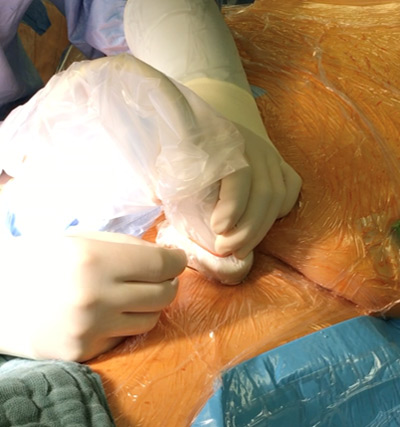
- Place the needle in the middle of the transducer and penetrate the skin (Figure 3).

Figure 3: Positioning of the ultrasound probe and the needle.
- Maintain visualization of the needle tip, which will require continuous adjustments of the probe and small movements of the needle (Video 3).
- Once the needle tip reaches the anterior vessel wall the artery may be punctured under visual control (Video 4).
Points of specific attention
Obese patients require a specific technique: the lower belly should be pushed upwards, and the puncture should be in a straight direction (green arrow in Figure 4), otherwise the insertion of the sheath or preclosure device may become difficult.
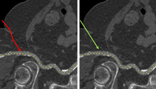
Figure 4: The red arrow depicts a puncture with slight changes of the angle, the green arrow indicates a straight puncture.
A word from Alexander Wolf
An excellent demonstration of how to perform ultrasound-guided femoral puncture. Very important in avoiding vascular complication, in particular for new TAVI Operators, who are nowadays predominantly trained on radial access for coronary interventions.


1 comment
Nice demonstration