198 results
Risk evaluation in asymptomatic patients: what is the current and future role of CT angiography?
21 May 2026 – From EuroPCR 2026
Ronak Rajani and Carlos Collet discuss the role of coronary CT angiography in cardiovascular risk assessment among asymptomatic individuals. They review current approaches to risk stratification and examine whether imaging can provide incremental value beyond traditional clinical risk factors.
The conversation explores the growing range of information available...
How the latest innovations in OCT help us improve PCI
20 May 2026 – From EuroPCR 2026
In this discussion, Thomas Johnson, Nieves Gonzalo and Simone Biscaglia explore the latest advances with Ultreon 3.0, a new platform that integrates OCT imaging and physiology into a single workflow for PCI guidance. They highlight how faster pullback and reduced contrast volume can expand access to intracoronary...
What is new in intracoronary physiology in 2026 and why does it matter?
19 May 2026 – From EuroPCR 2026
Javier Escaned and Niels Van Royen explore the evolving role of physiology in coronary assessment, with a focus on angiography-derived and imaging-derived indices, highlighting the growing potential of AI-driven tools to integrate anatomical and functional data, enabling more comprehensive and flexible decision-making without the need for...
How will the ALL‑RISE randomised trial evaluating angiography‑derived FFR impact our practice?
20 May 2026 – From EuroPCR 2026
Rasha Al-Lamee and Margaret Mcentegart review the results of the ALL‑RISE randomised trial, evaluating angiography-derived FFR (FFRangio) compared with wire-based physiology. The study demonstrated non-inferiority in clinical outcomes at one year, while offering significant advantages in terms of workflow, including reduced procedure time, fluoroscopy, and contrast...
How are the FRACTURE IDE trial results on laser-based intravascular lithotripsy going to impact the management of calcified lesions?
19 May 2026 – From EuroPCR 2026
Evald Christiansen and Margaret McEntegart review the results of the FRACTURE IDE trial which evaluated a novel optical laser-based intravascular lithotripsy (IVL) system for the treatment of severely calcified coronary lesions. The study demonstrated high rates of procedural success and safety, with low rates of clinically...
Is the plaque-biology assessment the future of imaging?
19 May 2026 – From EuroPCR 2026
Javier Escaned and Thomas Keeble discuss how near-infrared spectroscopy (NIRS) is emerging as a valuable tool to assess plaque biology in coronary arteries by measuring lipid content, a key marker of plaque vulnerability. Unlike traditional imaging that mainly describes structure, NIRS identifies high-risk, lipid-rich plaques using...
Calcium Skills Lab: EuroPCR 2026 highlights
19 May 2026 – From EuroPCR 2026
As the management of heavily calcified coronary lesions becomes increasingly complex, the need for practical, hands-on education continues to grow. At EuroPCR 2026, a brand-new initiative aims to address this challenge: the Calcium Skills Lab.
In this discussion, Angela McInerney and Mohamed Abdel-Wahab present this innovative programme...
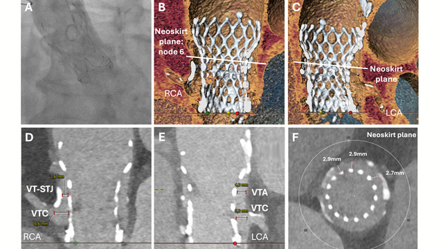
Rapid CT-guided emergency TAVI-in-TAVI-in-valve for cardiogenic shock with high coronary obstruction risk
14 Apr 2026
An 85-year-old man presented in cardiogenic shock with torrential aortic regurgitation after prior surgical and TAVI valve-in-valve procedures. With high coronary obstruction risk, urgent CT assessment and in-silico simulations guided a rapid redo TAVI-in-TAVI-in-valve implantation, demonstrating that CT-assisted emergency redo-TAVI is feasible and lifesaving in high-risk...
ALL-RISE: A large-scale, global randomized trial of coronary physiology derived from conventional angiography compared with an invasive pressure wire-based approach to guide PCI
28 Mar 2026
Mirvat Al Asnag provides her take on the ALL-RISE randomised trial presented by Ajay J. Kirtane at ACC.26 in New Orleans.

Author

IVUS-guided versus angiography-guided PCI in unprotected left main coronary artery disease – The OPTIMAL trial
31 Mar 2026
Ali Nazmi Calik provides his take on the results of the OPTIMAL trial presented by Luca Testa at ACC.26 in New Orleans.

Author