198 results
Imaging and physiology-guided DCB strategy
20 May 2026 – From EuroPCR 2026
Delve into advanced imaging and physiology-guided drug-coated balloon (DCB) strategies for complex coronary lesions. This session presents integrated approaches combining morphology and physiology, OCT guidance, and real-world cases highlighting DCB use in multivessel disease and calcified lesions.

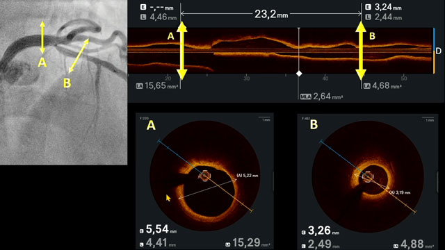
Intracoronary imaging to guided DCB PCI
20 May 2026 – From EuroPCR 2026
This session highlights the role of intracoronary imaging techniques such as OCT and IVUS in guiding drug-coated balloon (DCB) PCI. It explores novel imaging-guided algorithms for complex bifurcations, prediction of drug-eluting balloon failure, management of calcified nodules, re-entry into true lumen during CTO, and tackling aneurysms...
Imaging to the rescue in LAA closure
20 May 2026 – From EuroPCR 2026
This session highlights the pivotal role of advanced imaging modalities in left atrial appendage (LAA) closure. Topics include novel risk assessment workflows for air embolism, evaluation protocols using cardiac CT and 3D TEE, reproducibility of anatomical classifications, and long-term follow-up outcomes with various imaging techniques.

Choosing the right strategy for isolated ostial left anterior descending artery or circumflex coronary lesions
20 May 2026 – From EuroPCR 2026
This session examines optimal strategies for managing isolated ostial lesions in the left anterior descending and circumflex arteries, comparing left main crossover, ostial stenting, and drug-coated balloon (DCB) approaches. It emphasizes integrating multimodality imaging for informed decision-making, illustrated through detailed case discussions.
Typical chest pain: atypical angiogram
20 May 2026 – From EuroPCR 2026
This session addresses the management challenges posed by acute coronary syndromes when angiographic findings are ambiguous or atypical. It highlights the role of intravascular imaging in clarifying diagnoses and discusses recent insights from clinical cases and key studies such as DISCO and the latest NSTEMI EuroIntervention...
When the angiogram shows no answer for your AMI patient: what's next?
20 May 2026 – From EuroPCR 2026
This session focuses on non-occlusive causes of acute coronary syndromes (ACS) when angiograms do not reveal clear pathology. It emphasizes the diagnostic value of intracoronary imaging for conditions such as MINOCA and spontaneous coronary artery dissection (SCAD), and discusses optimal treatment strategies and prognostic considerations.
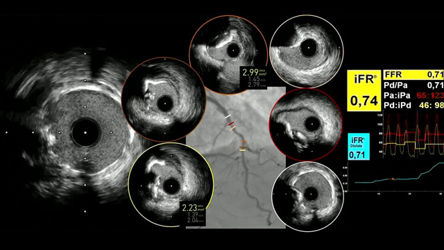
Imaging guided ultralow contrast PCI
20 May 2026 – From EuroPCR 2026
This session focuses on imaging-guided ultralow contrast PCI techniques designed to minimize contrast-induced nephropathy risk. It includes IVUS-guided strategies for distal left main bifurcation PCI, zero-contrast interventions in renal transplant patients, and precision stent placement without contrast use, emphasizing safety, precision, and frugality in contemporary PCI...
Intracoronary imaging guidance in challenging PCI scenarios
20 May 2026 – From EuroPCR 2026
This session highlights the critical role of intracoronary imaging, particularly IVUS, in guiding complex percutaneous coronary interventions. It presents techniques such as IVUS-guided puncture of neo-ostia, stent implantation for ostial lesions, and innovative procedural methods like the Heart POTTER technique, underscoring the importance of precise imaging...
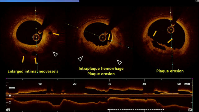
Intracoronary imaging insights in ACS
20 May 2026 – From EuroPCR 2026
Discover how intracoronary imaging advances understanding and management of acute coronary syndromes (ACS). This session explores high-definition intravascular ultrasound for PCI guidance, novel predictive metrics, and the impact of emerging factors like microplastics on plaque instability, alongside pathophysiological insights into layered plaques and coronary ectasia in...
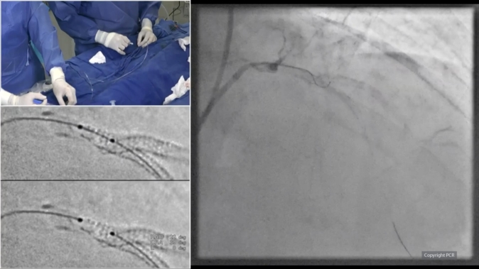
DK crush technique in a calcified LAD-diagonal bifurcation - LIVE case
Recommended by PCR
20 May 2026 – From EuroPCR 2026
A 53‑year‑old man with hypertension and dyslipidemia presented with an inferior MI treated with primary PCI of the RCA two months earlier. He was referred for staged treatment of a significant calcified LAD–diagonal bifurcation lesion. Lesion preparation included non‑compliant balloon dilatation, rotational atherectomy in the LAD...