06 Apr 2021
Femoral artery pseudoaneurysm expansion managed through subcutaneous fat embolization
Femoral artery pseudoaneurysm is a known complication following cardiac catheterization. Rarely, expansion of the pseudoaneurysm can occur and is often catastrophic. We describe a unique method employed by our centre to deal with such a complication.
This case was originally published on Twitter by @ezmanshariff via #CardioTwitterCase
Clinical presentation
We present the case of a 61-year-old woman who underwent percutaneous coronary intervention of her left anterior descending and left circumflex arteries via right femoral artery access. She suffered from diabetes, hypertension and end-stage renal failure and was on regular haemodialysis through a permacath. Following removal of her femoral sheath 12 hours earlier, she became increasingly unwell, recording a blood pressure of 7/30 mm Hg, pulse rate 120 beats/minute and Glasgow Coma Scale of 13 (Eyes 4, Verbal 4, Motor 5). We noted a large swelling and bruising of the right femoral (Figure 1-A). Urgent bedside ultrasonography was performed, which revealed a large femoral artery pseudoaneurysm (FAP) (Figure 1-B).
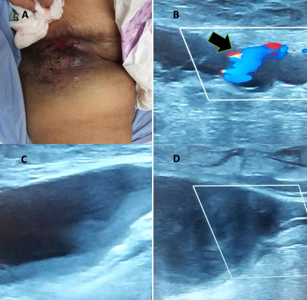
Figure 1: examination revealed left groin swelling and bruising, suggestive of a haematoma (A), and bedside ultrasound demonstrated pseudoaneurysm with evidence of progressive filling into the aneurysmal lumen via a ‘neck’ (black arrow) with the help of colour doppler imaging (B). Unfortunately, despite ultrasound-guided compression, there was progressive deterioration in the size of the pseudoaneurysm (C, D).
Case management
Alongside fluid resuscitation and commencing inotropic support, we had initially opted for ultrasound-guided vascular compression. Unfortunately, after 30 minutes, there was further evidence of FAP progression. Regrettably, we had no in-house vascular surgery services and despite a general surgical consult, the patient was deemed too unstable for transport to the operating theatre. Due to this situation, the decision was made to perform peripheral intervention to help tamponade the neck of the FAP.
Using left femoral artery access and a Judkins Right guiding catheter, angiography was performed on the right femoral artery and its tributaries, which revealed the location of the FAP neck (Video 1), before a 0.014 Sion Blue (Asahi) coronary wire was navigated down the right superficial femoral artery (SFA).
A 5.0/12 mm coronary balloon was initially inflated at various locations along the SFA, unsuccessfully. This was followed by inflating a combination of both a 5.0/12 mm coronary balloon down the SFA and a 4.5/15 mm one along the profunda femoris artery (PFA) after navigating an additional 0.014 Sion Blue wire down the PFA, to no avail (Video 2).
Unfortunately, there were no available balloons or grafts routinely used for peripheral interventions readily available at the time in our centre. There were also no coronary balloons larger than the ones of a 5.0 mm diameter.
As the patient’s condition continued to deteriorate, a decision was made to seal the neck of the FAP. Again, unluckily, we did not have either embolization coils or thrombin readily available. We decided to harvest subcutaneous fat from the patient’s abdomen, and form a solution using contrast, before introducing the mixture to the neck of the FAP through a Finecross microcatheter (Figure 2).
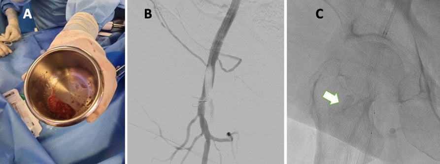
Figure 2: subcutaneous fat from the patient’s abdomen was harvested (A) and mized with contrast to be used for the purpose of embolization. A coronary wire was slowly guided down the neck of the pseudoaneurysm (B), before introducing a Finecross microcatheter (C) which was then used to deliver the ‘fat-contrast’ mixture to the neck (white arrow), allowing it to seal off.
This was performed alongside a 5.0/12 mm coronary balloon inflation, which was successful in both sealing the neck of the pseudoaneurysm and enabling the patient to be stabilized for the night (Video 3).
Original tweet and Twitter discussion
61y F DM, HTN, ESRD on permcath. PCI to LM-LAD and DEB to LCx performed 24h ago via femoral due to poor radial pulses. Hypotensive 12h post-removal of Rt femoral sheath. BP 70/30. HR 120. GCS 13/15. Noted tense in the RT femoral area. pic.twitter.com/18RH7WrYDl
— Ezman Shariff (@ezmanshariff) March 21, 2021
Author

2 comments
GOOD CASE .. !!!. HOW TO MAKE THE FAT AND CONTRAST MIX ...?
GOOD CASE .. !!!. HOW TO MAKE THE FAT AND CONTRAST MIX ...?