Calcified breast in the heart: which imaging modality is diagnostic? Invasive or non-invasive?
Image in interventional cardiovascular medicine: what is displayed on this image? ... And do not hesitate to comment on how you would treat this case!
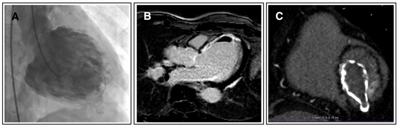
A 57-year-old smoker with a history of multivessel coronary artery disease (MVD) and healed spontaneous coronary artery dissection (SCAD) of the left anterior descending (LAD) presented with dyspnea and chest discomfort. During fluoroscopy, there was a shadow of a strange calcified area in the heart. Echocardiography showed severely reduced ejection fraction (EF = 20 %).
Six months later, the patient was admitted with non-ST Elevation Myocardial Infarction (NSTEMI). The new angiography showed no significant changes, but the calcified breast-like area with a nipple-like protrusion was still visible.
An image is worth a 1,000 words: participate in the quiz below to tell us what you see in this image!
Calcified breast in the heart: which imaging modality is diagnostic? Invasive or non-invasive?
Authors
Mehrpooya Maryam1, Babaei Mohammadreza1- Tehran University of Medical Science - School of Medicine-Imam Khomeini Hospital Complex, Tehran, IRAN
No comments yet!