What is it with that crumpled coronary: a lesion or a pseudo-lesion?
Image in interventional cardiovascular medicine: what is displayed on this image? ... And do not hesitate to comment on how you would treat this case!
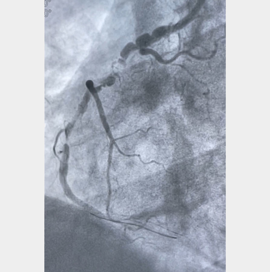
A 66-year-old male patient presented to hospital with a complaint of chest pain 1 hour prior to admission. A diagnosis of acute inferior wall myocardial infarction was made, and he was thrombolysed with Tenecteplase.
A coronary angiography was performed, which showed a very tortuous RCA vessel with a tight lesion (about 90 %) in mid-RCA (right coronary artery), with a post-stenotic aneurysm. PTCA to RCA was planned. During the procedure, as soon as the 0.014 inch BMW (balance Middleweight Wire) was advanced, it could not cross the tortuous proximal part of the vessel. An 0.014 inch All Star wire was taken and advanced into the distal end of the vessel with the support of a 2 mm*12 mm Mini Trek balloon. As soon as the wire crossed the lesion, the proximal part of the patient’s artery showed increased narrowing. The ECG on the monitor at this time showed ST elevation and the rate dropped to 40 beats / minute. The balloon was quickly inflated at the site of the lesion. Thereafter, the vessel had a slow flow distally. IV nitroglycerin was repeated and 2.5 mm*23 mm Everolimus stent was deployed at the site of the lesion. Post-procedure, the vessel had TIMI 3 flow with BP of 90 / 60 mm. ECG showed settling of ST elevation in inferior leads. Patient was relieved of his chest pain and transferred out of the cathlab.
An image is worth a 1,000 words: participate in the quiz below to tell us what you see in this image!
What is it with that crumpled coronary: a lesion or a pseudo-lesion?
Authors
Ruhani Bali1, Rajneesh Calton1
- Deepak Heart Institute - Best Heart Specialist Hospital | Cardiologist in Ludhiana, Punjab, INDIA
2 comments
Recognition of this effect will help us to avoid unnecessary stenting.
This effect disappear after remove coronary wier and given inj NTG or nikoran intracoronary in this RCA proximal area concertina but mid area 99 percent disease