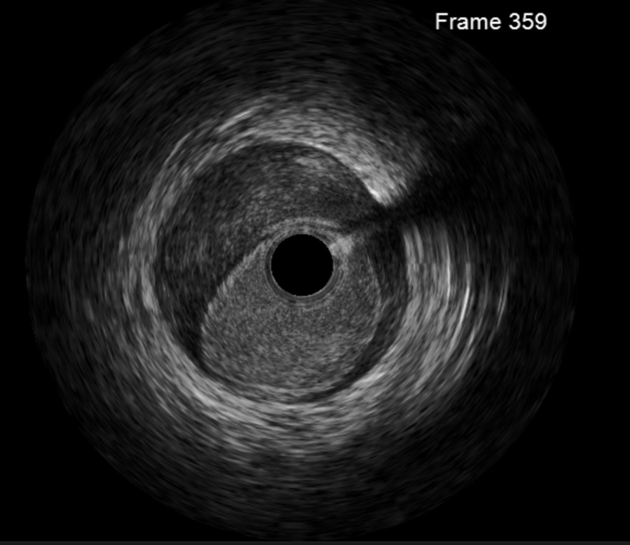
Intravascular ultrasound (IVUS) of the LAD
Image in interventional cardiovascular medicine: what is displayed on this image? ... And do not hesitate to comment on how you would treat this case!
A 50-year-old woman with no significant cardiovascular risk factors underwent open reduction and internal fixation (ORIF) for an ankle fracture.
Two days after surgery, while recovering on the ward, she developed sudden-onset central chest pain with ECG changes suggestive of anterior wall ischaemia.
Coronary angiography demonstrated:
- distal LAD occlusion
- diffuse narrowing of the proximal and mid LAD
An image is worth a 1,000 words: participate in the quiz below to tell us what you see in this image!
Intravascular ultrasound (IVUS) of the LAD
Author
Reddy K.1, Toor I. S.1
- Manchester Royal Infirmary, Manchester, UNITED KINGDOM
No comments yet!