Where is the lesion ?
Image in interventional cardiovascular medicine: what is displayed on this image? ... And do not hesitate to comment on how you would treat this case!
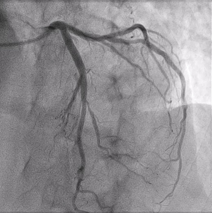
A 60-year-old male patient with no known underlying disease presented with chest pain at rest persisting for two days without resolution.
ECG showed ST-segment depression in leads II, III, and aVF.
Troponin T increased from 150 to 550.
Left coronary angiogram in LAO cranial view shown in the image.
An image is worth a 1,000 words: participate in the quiz below to tell us what you see in this image!
Where is the lesion ?
Author
Kieattisaksopon Suppavich1
- Somdech Phra Pinklao Hospital, Bangkok, THAILAND
1 comment
Spider view sometimes make it more clear.