Mysterious coronary artery
Image in interventional cardiovascular medicine: what is displayed on this image? ... And do not hesitate to comment on how you would treat this case!
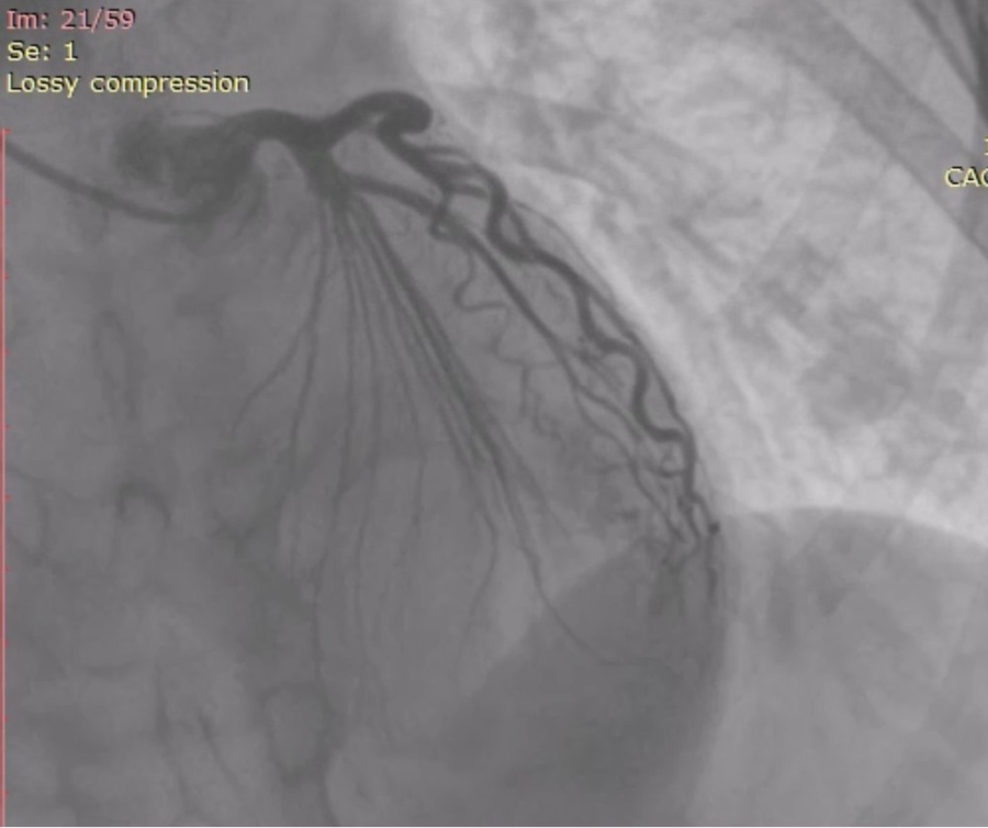
A 56-year-old hypertensive female was admitted to our hospital with complaints of dyspnoea on exertion and retrosternal chest pain. Physical examination and laboratory screening results were normal. Twelve lead electrocardiogram revealed LVH and LV strain pattern. Echocardiogram revealed concentric left ventricular hypertrophy, LVEF was 60%. The patient was transferred for coronary angiography.
An image is worth a 1,000 words: participate in the quiz below to tell us what you see in this image!
Mysterious coronary artery
Author
Joshi P.1
- Priyanka Hospital & Cardiac Centre (PHCC), Rajasthan, INDIA
No comments yet!