OCT image of bifurcation lesion
Image in interventional cardiovascular medicine: what is displayed on this image? ... And do not hesitate to comment on how you would treat this case!
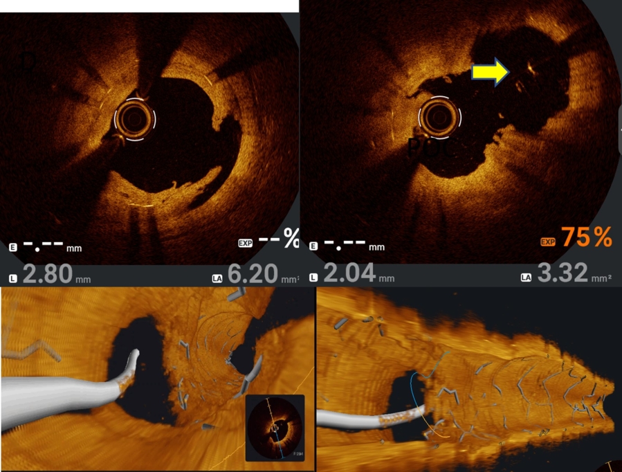
A 54-year-old female with risk factors: Hypertension, type diabetes mellitus, H/o PTCA to LAD/D1 bifurcation lesion - TAP stenting in 2020, presented with chest pain at rest. Vitals ok, ECG ST, t changes in precordial leads, 2d echo mild LV dysfunction (LVEF 48%).
CAG revealed LAD/D1 ISR, after a heart team discussion patient was planned for PTCA to LAD/D1 bifurcation PCI. Transradial route - Ebu 7f guide catheter, LAD could not be wired, D1 stent protrusion in LAD, so D1 was wired and D1 stent was dilated with 3mm NC balloon, TAP was converted to Cuolette, then LAD was wired through stent struts and dilation was iodine with 2.5mmm balloon and 3mm cutting balloon, then kissing balloon dilation was done and sequential 2.5X20mm DCB in D1 and 3x25mm DCB was done in LAD with good end results.
An image is worth a 1,000 words: participate in the quiz below to tell us what you see in this image!
OCT image of bifurcation lesion
Authors
Preeti Sharma1
- Max superspeciality hospital, Dehradun, INDIA
No comments yet!