The phantom branch from the left main coronary artery
Image in interventional cardiovascular medicine: what is displayed on this image? ... And do not hesitate to comment on how you would treat this case!
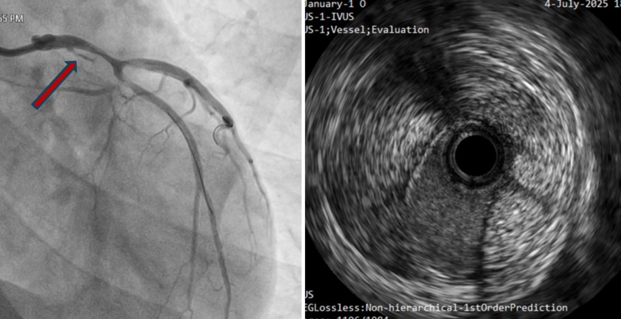
A 48-year-old male patient with marfanoid habitus presented with acute-onset chest pain 5 days prior. ECG was suggestive of an evolved lateral wall myocardial infarction, and echocardiography showed regional wall motion abnormalities (RWMA) in the left circumflex territory.
An image is worth a 1,000 words: participate in the quiz below to tell us what you see in this image!
The phantom branch from the left main coronary artery
Author
Biswas A.1
- U N Mehta Institute of Cardiology & Research Centre, Gujarat, INDIA
No comments yet!