20 May 2025
Complex PCI in calcific coronary artery disease - LIVE Case
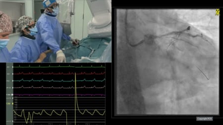
Calcified RCA lesion PCI with OCT-guided lesion preparation
Summary
A 76-year-old male patient presented with a significant history of coronary artery disease: CABG with LIMA/LAD and RIMA/OM (1996), RCA PCI with BVS (2016), OM PCI (2017), RIMA occlusion and OM retenosis treated with DCB (2022) with preserved LV. He was admitted for pre-operative evaluation prior to surgical carotid endarterectomy. He also presented with a stable angina (CCS 2). Coronary angiography revealed a calcified RCA stenosis on the BVS site.
Following extensive lesion preparation using orbital atherectomy and non-compliant balloon dilation guided by OCT, three stents were successfully implanted.
LIVE Educational Case from Clinique Pasteur - Toulouse, France
Navigate the video by moving your mouse over the chapter icon in the toolbar

Key moments
- 5:38-7:34 - Imaging analysis
- 17:10-20:01- Orbital atherectomy
- 42:56-44:10 - OCT evaluation
- 1:10:56-1:15:57 - Procedural analysis
Keywords: RCA PCI - Orbital atherectomy - OCT - IVUS


