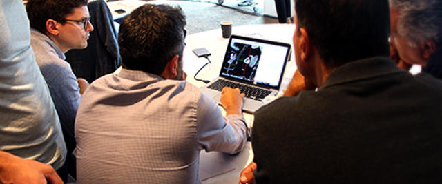
PCR London Valves 2018 - Structural imaging workshops
“The Eyes of the Heart Team” - Course Director Nicolo Piazza shares on the new implementation of an innovative Imaging Learning Centre
Unlike cardiac surgeons who have direct visualization of heart structures, interventional cardiologists are dependent upon cardiovascular imaging to organize and conduct their procedures. An integrative approach to cardiovascular imaging using fluoroscopy, echocardiography and multi-slice computed tomography is critical in the success of structural heart disease interventions.
As a consequence, those interested in performing transcatheter structural heart procedures are to become “interventional imaging experts”. In addition to the potential lack of knowledge and skills in cardiovascular imaging, there is an equal and possibly greater peril in the lack of awareness of its importance and practicalities. Interventional cardiology training programs should provide a structured approach to learning cardiovascular imaging with a defined set of objectives.
The knowledge and skill sets required by the “interventional imaging expert” span across the spectrum of image acquisition, analysis, interpretation and application. Furthermore, correlation across imaging modalities is key to simplifying and understanding the so-called “complexities” of cardiovascular imaging. For instance, realizing that the left heart can be viewed in a 2-, 3- and short-chambered axis, irrespective of the imaging modality, simplifies the required knowledge for identification of anatomical structures across imaging modalities.
Developing a common language between the operator and non-invasive imaging expert can be challenging. While MSCT and fluoroscopy is more accustomed to using the attitudinal description of the heart to localise structures (i.e. superior/inferior, anterior/posterior, right/left), echocardiography uses terms such as medial, lateral and septal that can confuse the operator who is navigating according to MSCT/fluoroscopic information. The attitudinal description of the heart can be assumed across imaging modalities and can simplify communication. Both papillary muscles, for example, can be identified as posterior structures oriented in a superior and inferior orientation, respectively.
The first step in becoming an “interventional imaging expert” requires a thorough understanding of the attitudinal description of cardiac structures. This knowledge can be acquired via didactic lectures which is then extended to the wet lab while analysing human or animal heart specimens. Subsequently, the anatomical structures can be identified and localised according to chamber anatomy (2-, 3- and short-axis views), allowing physicians to bridge this knowledge across imaging modalities. The mastery of basic anatomy and imaging can then be applied to various transcatheter heart procedures. Following this knowledge acquisition, imaging skills will be developed through imaging workshops.
At the end of the course, we hope that attendees will be able to understand the correlation between imaging modalities, use multi-slice computed tomography as a central tool to better understand fluoroscopic and echocardiographic imaging, and apply the newly acquired knowledge and skills sets towards improving patient outcomes.
