30 May 2023
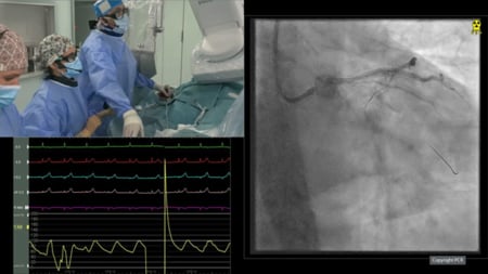
Severely retroflexed calcified circumflex artery – Tackled with left distal radial approach!
#CardioTwitterCase originally published on Twitter
Calcified, retroflexed subtotal long occlusion of left circumflex into OM1 successfully intervened after lithotripsy via left distal radial (anatomical) snuffbox approach
This case was originally published on Twitter by @pras_senmd
Case description
Distal radial access has been increasingly used for multiple reasons, including ease of hemostasis, patient comfort, potential for decreased incidence of spasm. However, recent literature suggests that radial artery occlusion rates might be similar compared to conventional radial access. Nevertheless, left distal radial access must be strongly considered for operator and patient convenience in patients with bypass surgery involving the left internal mammary artery.
We present a 63-year-old male patient with history of CAD s/p three-vessel CABG (LIMA to LAD, SVG to PDA and OM1) in 2020, type 2 diabetes mellitus, hypertension, hyperlipidemia, obesity s/p gastric bypass in 2009, who presented with unstable angina symptoms, and who underwent complex intervention for a severely calcified tortuous retroflexed left circumflex artery into OM1, that had subtotal long occlusion.
After ultrasound guided left distal radial access, and completion of diagnostic angiography, decision was made to intervene on the left circumflex to OM1 branch that had a long calcified severely diseased segment. A 6Fr EBU 3.5 guide catheter was used, and a runthrough coronary wire, with the help of a teleport microcatheter, was used to cross the LCx/OM lesion and parked distally in OM. Initially, we had difficulty advancing a 1.0 mm compliant ballon across the lesion. We then used a 5.5F Guideliner, and were able to advance the 1.0 x 8 mm compliant balloon.
This was followed by a 1.5 x 8 mm compliant balloon and, then, a 2.0 x 15 mm compliant balloon. IVUS showed severely calcified vessel, with nearly 360 degree arc of calcium. Hence, decision was made to perform intravascular lithotripsy with a Shockwave 2.5 mm balloon (distal vessel 1 :1 sizing). All 80 pulses were delivered. Adequate expansion was confirmed.
Then, we proceeded with a Synergy XD 2.5 mm x 38 mm DES in the proximal LCX extending to the OM branch followed by an overlapping a Synergy XD 3.0 x 8 mm.
Post-dilation was performed with a high-pressure inflation, using the 2.5 mm NC balloon, and a 3.0 mm NC balloon in the distal proximal portions respectively, with excellent angiographic result (See videos below). Pressure dressing was applied for hemostasis, it was removed after 2 hours, and patient was discharged within 24 hours.
Videos
Final remarks
Contrary to traditional belief that tortuous, calcified, retroflexed arteries need more support, and that attempting PCI via femoral route may be better, we show that minimally invasive left distal anatomic snuffbox approach is a much safer and more effective way with modern PCI tools such as microcatheters and guide extensions, and, importantly, intravascular lithotripsy.
Original tweet and Twitter discussion
Unstable Angina in a 60 y/o
— Prasanna Sengodan (@pras_senMD) May 1, 2023
Patent LIMA-LAD, patent SVG- Ramus, PCI to LCx-OM1 (not grafted)
Quite tortuous, calcified LCx-OM1.
Left distal radial access
Microcatheter, Guide extension, IVL to get the job done. @RameshDaggubati@Akram_Kawsara@vikjag45@ShockwaveIVLpic.twitter.com/1HjG4msFFU


1 comment
Why right distal radial artery not used ; if used which size guider can used. It’s great case and well executed with latest tools.