16 May 2024
Complex coronary artery disease: how to use intracoronary imaging in daily practice challenges?
With the collaboration of the Korean Society of Interventional Cardiology (KSIC), Interventional Cardiology Association of the Spanish Society of Cardiology and Cardiac Society of Australia and New Zealand - Interventional Council
Summary
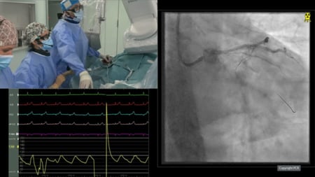
This session, in collaboration with various cardiology societies, focuses on the use of intracoronary imaging to optimize percutaneous coronary intervention (PCI) in complex coronary artery disease. It presents case studies demonstrating the role of imaging-based assessment of coronary calcified lesions, the importance of a standardized imaging workflow, and how intracoronary imaging can guide decision-making and improve procedural outcomes.
Learning Objectives
- To learn how to optimise PCI using intracoronary imaging
- To understand imaging-based assessment of coronary calcified lesions
- To clarify the role of a standardised imaging workflow in PCI


