71 results
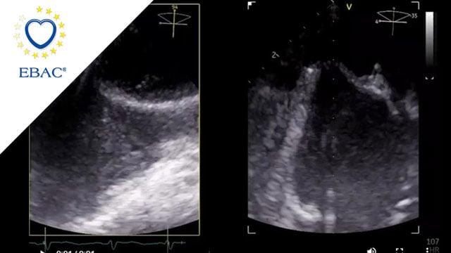
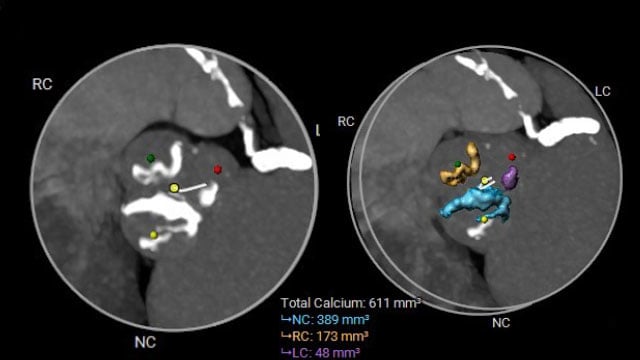
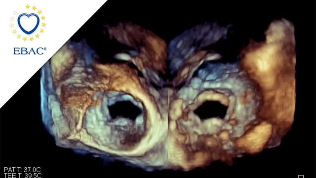
Very small LAA with a challenging closure
15 Jul 2026
Recurrent strokes despite anticoagulation, a failed first attempt at left atrial appendage closure (LAAC), and a challenging small chicken-wing anatomy: this case brings together several factors that complicate procedural planning and device selection.
In a 65-year-old woman with paroxysmal atrial fibrillation, pre-procedural TOE and CT imaging highlighted...

Author
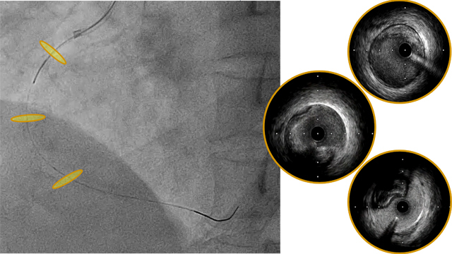
An unconventional pathway to the management of an iatrogenic dissection
23 Jun 2026
Iatrogenic coronary dissection is a rare but potentially life-threatening complication of PCI. Its management can be challenging, particularly when antegrade wiring fails and flow is compromised, requiring escalation to advanced bailout strategies.

Author
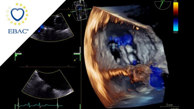
Making the complex simple: bicaval valve implantation as a rescue strategy
16 Jun 2026
An 80-year-old woman was referred for persistent right heart failure despite previous surgical mitral repair and subsequent transcatheter tricuspid intervention. Over time, she developed progressive symptoms with recurrent severe tricuspid regurgitation, refractory to optimised medical therapy.
Multimodality imaging confirmed significant right-sided chamber dilatation and complex valve anatomy,...

Author

Author

Author
From assessment to strategy: choosing the optimal treatment for severe tricuspid regurgitation
12 May 2026
Severe tricuspid regurgitation is no longer a bystander disease.
In this elderly patient with persistent symptoms despite optimised medical therapy, a comprehensive multimodality assessment becomes key to determining the right timing and the most appropriate transcatheter strategy.

Author

Author
High calcium, high stakes: managing severe AS with complex multivessel disease
12 May 2026
A 69-year-old woman with severe symptomatic aortic stenosis (NYHA III) and complex comorbidities, including three-vessel coronary artery disease, advanced COPD, and peripheral artery disease, presents with progressive dyspnoea despite recent PCI. How would you treat this patient?

Author

Author

Author
Transcatheter tricuspid valve replacement in carcinoid heart disease: A multimodality‑guided, patient-centered approach
17 Apr 2026
A 74-year-old woman with carcinoid syndrome develops progressive NYHA III dyspnoea and marked fatigue, despite well-controlled systemic symptoms. Echocardiography reveals torrential tricuspid regurgitation with carcinoid valve involvement, while surgical risk is deemed high due to age, frailty, and systemic disease. How would your Heart Team approach this...

Author
The future of aortic valve replacement - From index procedure to lifetime management
28 Apr 2026
A 68-year-old woman presents with symptomatic aortic stenosis and tricuspid valve anatomy, with no significant comorbidities and no evidence of coronary artery disease. Given her strong preference for a less invasive strategy, the Heart Team considers a transcatheter approach.
How would you treat this patient—and in case...

Author

Author

Author
A patient with severe MR and torrential TR
26 May 2026
This case explores the management of severe mitral regurgitation and torrential tricuspid regurgitation in a 71-year-old patient with a complex cardiovascular history.

Author
Mixed mitral valve disease in an irradiated chest with eggshell-like left atrium
27 Feb 2026
Managing mixed mitral valve disease is particularly challenging in patients with prior chest irradiation and extensive calcification.
A 40-year-old man presents with severe symptomatic MR, renal impairment, and an unusual eggshell-like left atrium. Imaging reveals complex mitral anatomy and critical constraints for transcatheter therapy.
How would you approach...

Author
When a surgical valve needs a second life
27 Feb 2026
A 79-year-old patient previously treated with surgical aortic valve replacement was referred after an episode of acute cardiac decompensation. Although clinically stabilised, further evaluation revealed degeneration of the bioprosthesis, and mixed aortic valve pathology.
This case focuses on the diagnostic assessment and the strategic considerations guiding the...

Author
