62 results
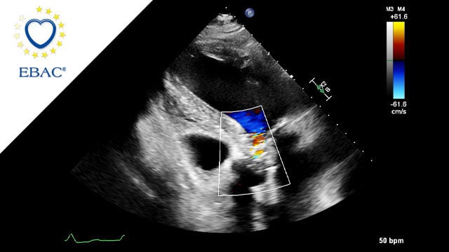
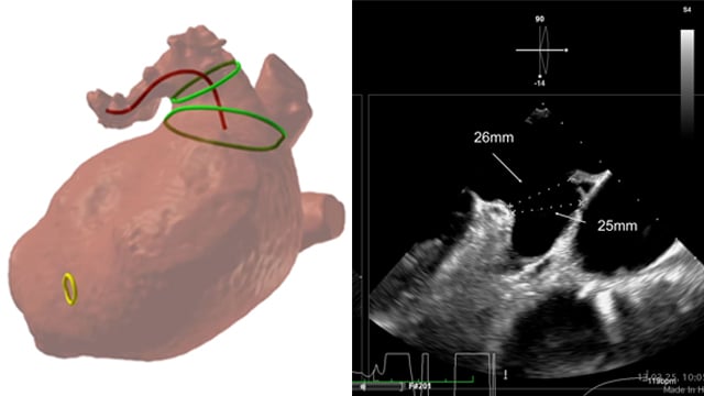
M-TEER with Mitraclip Gen5
03 Feb 2026
An 83-year-old female patient with HFrEF due to NICM and a history of arterial hypertension and persistent atrial fibrillation was referred with worsening exertional dyspnea, severe functional mitral regurgitation and moderate tricuspid regurgitation.
How would you treat this patient?

Author
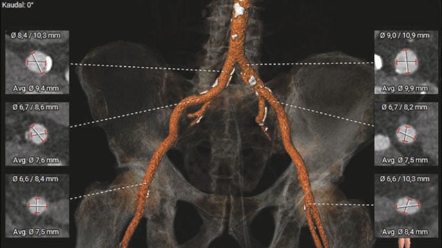
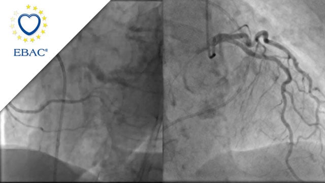
A challenging valve-in-valve scenario in a patient with prior aortic coarctation repair
02 Feb 2026
This case features a 79-year-old patient with severe aortic stenosis and a degenerated surgical bioprosthesis, who previously underwent complex aortic surgery for coarctation.
Redo surgery was high-risk, so a transcatheter valve-in-valve procedure was chosen.
Explore the case to see how anatomy, prosthesis sizing, and vascular access options guide...

Author

Author

Author

Author

Author

Mixed mitral valve disease in an irradiated chest with eggshell-like left atrium
26 Jan 2026
Managing mixed mitral valve disease is particularly challenging in patients with prior chest irradiation and extensive calcification.
A 40-year-old man presents with severe symptomatic MR, renal impairment, and an unusual eggshell-like left atrium. Imaging reveals complex mitral anatomy and critical constraints for transcatheter therapy.
How would you approach...

Author
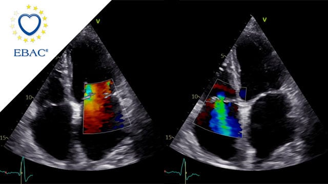
How should I treat a torrential tricuspid valve regurgitation in a young patient who remains symptomatic despite optimal medical therapy?
25 Dec 2025
A frail patient presents with worsening NYHA class III dyspnoea and peripheral oedema despite optimal medical therapy. Previously managed conservatively, severe tricuspid regurgitation has progressed to a torrential grade, with right-sided chamber dilatation and complex valve anatomy.
How would you treat this patient?

Author

Author
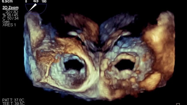
LAA closure with CT planning: challenging inferior chicken wing morphology
24 Dec 2025
This 77-year-old woman with paroxysmal AF and prior GI bleeding is referred for LAA closure, but CT reveals a shallow, inferior chicken-wing morphology that complicates device selection and planning. How would your Heart Team approach this challenging case?

Author
When a surgical valve needs a second life
23 Dec 2025
A 79-year-old patient previously treated with surgical aortic valve replacement was referred after an episode of acute cardiac decompensation. Although clinically stabilised, further evaluation revealed degeneration of the bioprosthesis, and mixed aortic valve pathology.
This case focuses on the diagnostic assessment and the strategic considerations guiding the...

Author
Severe mitral regurgitation in a patient at high risk for surgery with a suboptimal anatomy for M-TEER: what other options can we find?
04 Feb 2026
A 78-year-old male presents with recurrent hospitalisations for HFpEF and severe degenerative MR. Multimodality imaging reveals fibro-calcific leaflet disease, restricted motion, and a short posterior leaflet, making him a suboptimal candidate for M-TEER and prompting consideration of transcatheter mitral valve replacement. How would you treat?

Author

Author
Author
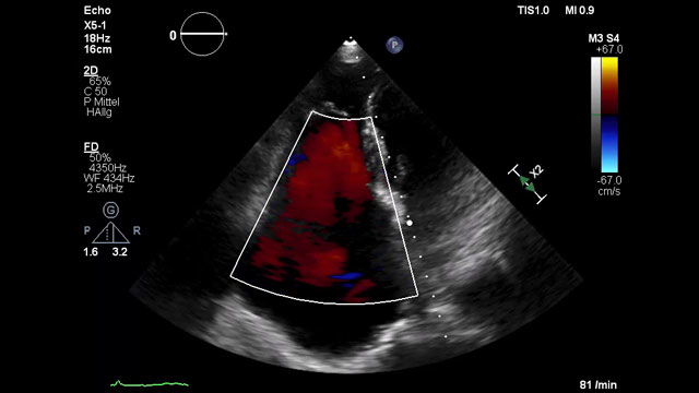
Impact of guideline-directed medical therapy on long-term outcome after tricuspid TEER
29 Jan 2026
A 79-year-old woman with advanced right heart failure (NYHA III) was referred after repeated hospitalisations for decompensation despite optimal medical therapy. Echocardiography revealed torrential functional tricuspid regurgitation with suitable anatomy for T-TEER. Given her high surgical risk (TRISCORE 7/12) and borderline pulmonary hypertension, how would you...

Author

Author
New roads begin where old ones end: a TAVI case
12 Jan 2026
An 85-year-old woman presents with exertional dyspnea and angina, alongside severe paradoxical low-flow, low-gradient aortic stenosis, marked mitral and tricuspid valve disease, and two-vessel coronary artery disease. Frailty markers and hostile pelvic anatomy further complicate her profile.
How would your Heart Team approach this challenging case?

Author

Author
When the first TAVI fails: no panic, just planning
19 Jan 2026
An 81-year-old man with history of severe aortic stenosis previously treated with a balloon-expandable transcatheter valve was referred for progressive exertional dyspnea due to bioprosthetic valve degeneration.
How would you treat this patient?

Author