Delayed compromise of a “super dominant” RCA and Shock
#CardioTwitterCase originally published on Twitter
Coronary obstruction is a known procedural complication of aortic valve replacement. However, delayed presentation few months after surgical AVR is a relatively unusual phenomenon. We describe a 75 year old woman with surgical AVR who presented with acute coronary syndrome and severe RV failure few months after the surgery.
These cases were originally published on Twitter by @TTelila
Clinical presentation
This is the case of a 75 year old woman who presented with severe right ventricular failure, congestive hepatopathy (severely elevated liver enzymes and coagulopathy with INR of 7) and rapid atrial fibrillation.
Case management
After initial hemodynamic stabilization and volume optimization, she was urgently taken to the cardiac catheterization laboratory to define her coronary anatomy and asses hemodynamics.
Right heart catheterization revealed a severely elevated biventricular filling pressures along and RV shock with PAPI=0.6.
Coronary angiography revealed a critical stenosis of the ostium of a “supper dominant” right coronary artery. The ostium of the RCA was in a very close proximity to the bioprosthetic aortic valve frame (25mm Inspiris). However, it was difficult to ascertain a clear mechanical compression.
Intravascular ultrasound revealed a large caliber RCA (5mm) with short segment of calcified plaque at the aorto-ostium. Given the clinical presentation, we performed a successful percutaneous Conroy intervention to the ostial RCA (Video 1) with IVUS guidance.
We used the Ostial flash balloon to flare the ostium (Figure 1).
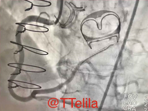
Figure 1. a Severe Ostial stenosis of a dominant RCA adjacent to the bioprosthetic aortic valve frame.
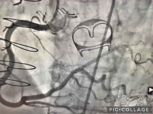
Figure 1. b Successful percutaneous coronary intervention to the Ostial RCA with implantation of a 5mm drug eluting stent.
Final remarks
Next day, patient was off inotropic support and few days later she was discharged home with marked clinical and biochemical improvement.
Original tweet and Twitter discussion
QUESTION: Has anyone seen this before —months after AVR? Pathophysiology? Management?
— Tesfaye A. Telila MD, FACC,FSCAI. (@TTelila) July 31, 2021
➡️Few months post AVR/MVR- 21mm Inspirus (AVR): presented in shock, massive fluid overload, RV failure, LFTs >3K, INR 7, severe TR & AFib-RVR ...@djc795@mmamas1973@SVRaoMD@DLBHATTMDpic.twitter.com/G2iAB9iNNb
Author

No comments yet!