19 Dec 2024
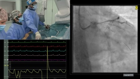
Undilatable calcified lesion - LIVE case
LAD calcified lesion PCI prepared by rotablator, cutting balloon and IVL
Summary
A 52-year-old male presenting with angina (CCS II) and normal LV function. Angiography revealed a long, severely calcified lesion in the proximal and mid-LAD, which was undilatable during a previous PCI.
The lesion was prepared using Rotablator (1.5 Burr), cutting balloon, Shockwave, and NC balloon, guided by OCT and IVUS. Two stents were implanted in the proximal and mid-LAD, while two drug-coated balloons (DCB) were used in the mid-distal and distal LAD, considering the patient’s young age and potential need for future CABG.
LIVE Educational Case from National Heart Centre at Royal Hospital - Muscat, Oman
Navigate the video by moving your mouse over the chapter icon in the toolbar

Key moments
- 12:00-13:43 - Imaging analysis
- 29:15-1:25:22 - Step-by-step preparation
- 29:15-36:00 - Rotablator procedure
Keywords: Calcified lesion, rotablator, IVL, Cutting balloon, DCB, OCT, IVUS, guideliner
Learning Objectives
- To learn how to use imaging to guide therapy in calcified lesions
- To learn how to select devices for plaque modification
- To learn how to optimise PCI results in heavily calcified lesions


