Aortic leaflets
Anatomy of the Aortic Valvar Complex
The normal aortic valve is trifoliate, the three leaflets form the aortic valve and provide its main sealing mechanism.
The normal aortic valve is trifoliate (Figure 5, Figure 6). Each of the three leaflets has a semilunar attachment within the aortic root and a free margin for coaptation with the other leaflets. At the level of the sinotubular junction, the semilunar hinges of adjacent leaflets come together to form the so-called commissures. If used literally, however, a "commissure” is a zone of apposition between adjacent structures. The true "commissures” within the valvar root, therefore, are the three zones of apposition between the leaflets extending from the so-called "commissures" to the valvar centroid.
The leaflets themselves are slightly thicker towards their free margins. Interindividual and intraindividual variability with respect to the width and height of the leaflets is common12.

Figure 5. Normal trileaflet aortic valvar leaflets
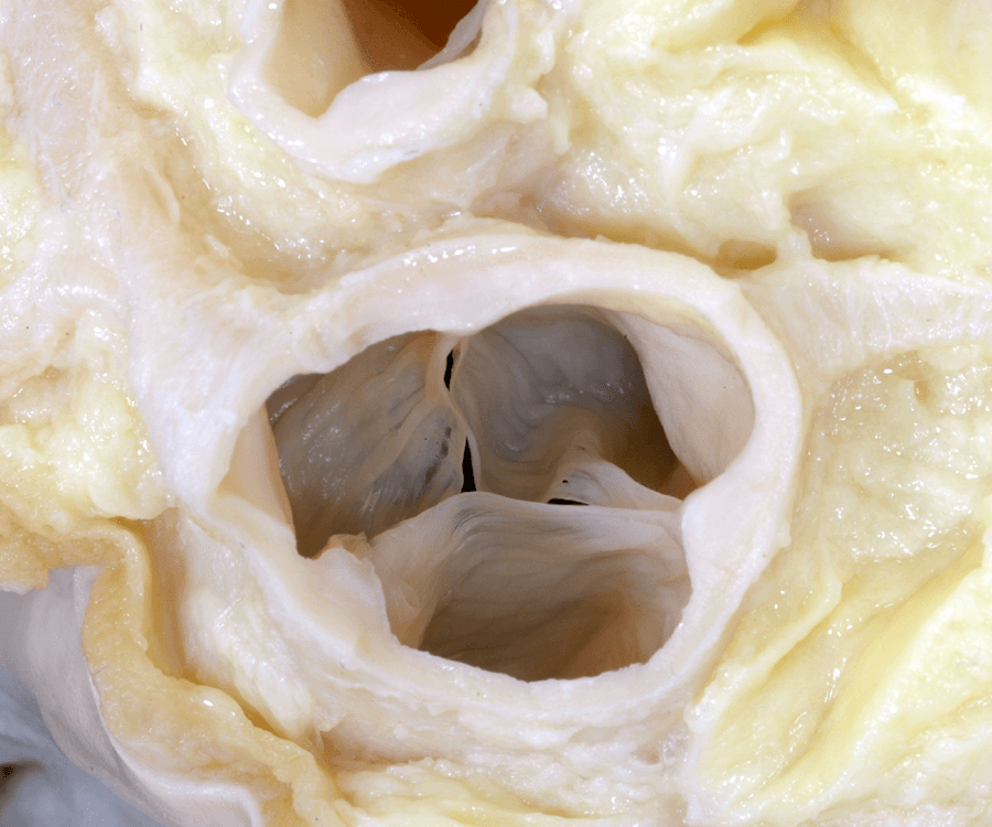
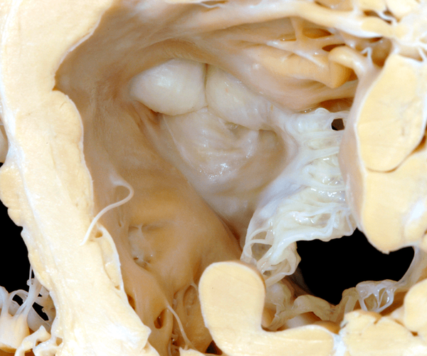
Figure 6. Normal trileaflet aortic valve leaflets


Figure 7. Dimensions of aortic leaflets
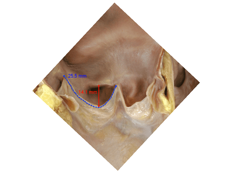
The average width, measured between the peripheral zones of attachment along the sinus ridge, is 25.5mm (Figure 7). The average height, measured from the base of the centre of the leaflet to its free edge, is 14.1mm (Figure 7). Coaptation along the zones of apposition occurs on the ventricular aspect of the leaflets, and involves the entire length of the free margin, taking place along approximately one-third of the total leaflet depth.
The central point of coaptation is thickened and is known as the lunule. The leaflets comprise a fibrous core and underlying subendothelial fibroelastic layers. In the setting of severe aortic stenosis, the leaflets become thickened, heavily calcified, and non-compliant, with a resultant reduction in the orificial area for the systolic ejection of blood by the left ventricle (Figure 8) and (Figure 9).

Figure 8. Calcific aortic stenosis
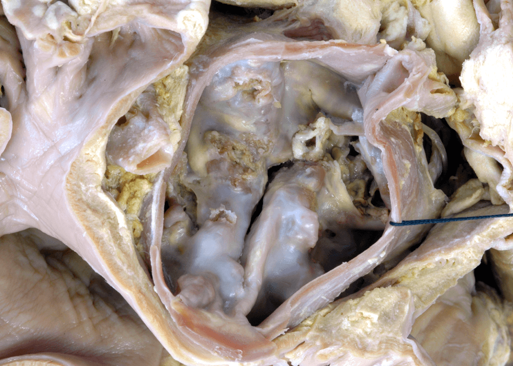
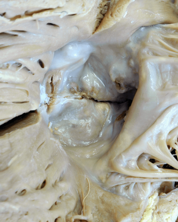
Figure 9. Calcific aortic stenosis

Normal trileaflet aortic valve leaflets
This close-up, short-axis view from the base of the heart shows the normal aortic valve with two of the leaflets and aortic sinuses adjacent to the pulmonary trunk, the right (red star) aortic sinus typically giving rise to the right coronary artery and the left (blue star) aortic sinus the left coronary artery.
The non-adjacent aortic sinus is represented by the yellow star. The zones of apposition (red arrows) between the leaflets extend from their attachments at the sinotubular junction (black stars) to the centre of the valvar orifice.
Historically, it was only the peripheral attachments which were referred to as the commissures. Actually, it is the entirety of the zone of apposition that is the commissure.
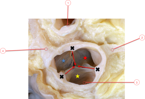
Figure 5. Normal trileaflet aortic valvar leaflets

1 - Pulmonary trunk
2 - Right coronary artery
3 - Aortic valve
4 - Left coronary artery
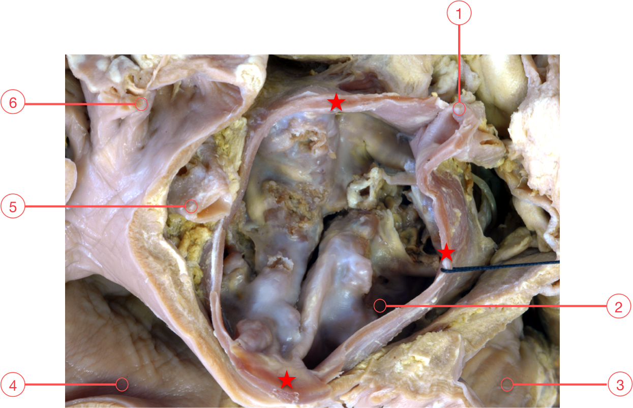
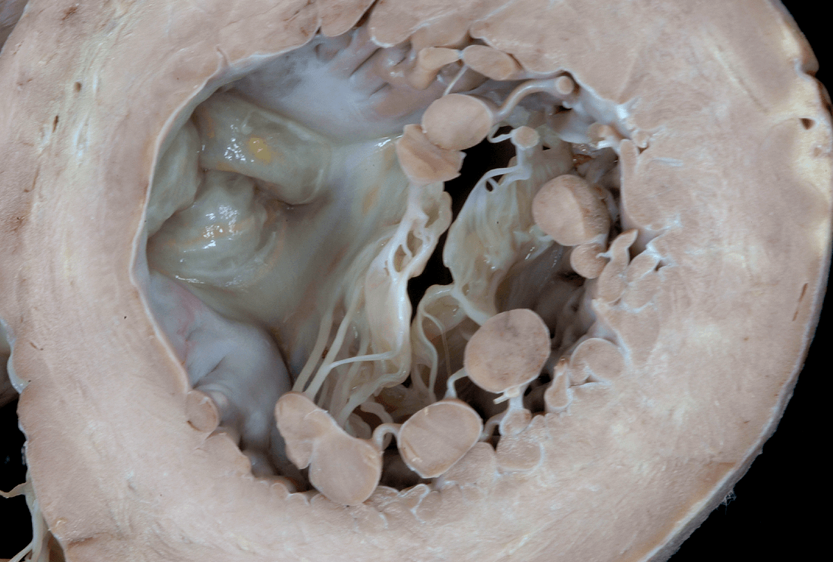
The aortic to mitral fibrous continuity (black dots) and the aortic valve are viewed from the apex of the left ventricle. The mitral valve is supported by tendinous cords arising from the paired papillary muscles with no attachments to the ventricular septum.
Within the left ventricle, the aortic root interposes between the mitral valve and the interventricular septum.
The left ventricular outlet has a partially fibrous and partially muscular wall. Two of the leaflets which guard the outlet or aortic root are in fibrous continuity with the anterior or aortic leaflet of the mitral valve. These are the non-adjacent (yellow star) and the left (blue star) facing or left coronary leaflet with the third leaflet, the right (red star) facing or right coronary leaflet.
The right and left fibrous trigones form the two ends of this fibrous continuity. The aortic root and mitral valve are anchored within the roof of the left ventricle where the fibrous trigones attach to the crest of the muscular ventricular septum. The fibrous trigones are thickened areas within the aortic to mitral fibrous continuity and the right fibrous trigone is continuous with the membranous septum. The right fibrous trigone and the membranous septum together form the central fibrous body.

Figure 6. Normal trileaflet aortic valve leaflets
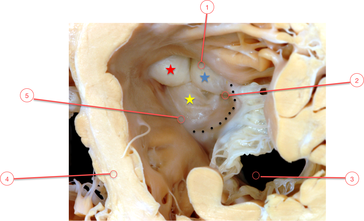
1 - Aortic Valve
2 - Left fibrous trigone
3 - Mitral valve
4 - Interventricular septum
5 - Right fibrous trigone
Dimensions of aortic leaflets
The average length and height of the aortic valve leaflets are 25.5 and 14.1mm, respectively.
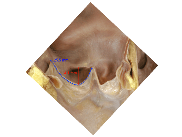
Figure 7. Dimensions of aortic leaflets
Calcific aortic stenosis
The aortic root is viewed from the base of the heart and has been transected just below the sinotubular junction along the most anterior aspect. The aortic valvar leaflets are extremely thickened and are entirely calcified. The leaflets are non-compliant with only a small, slit-like, eccentric opening.
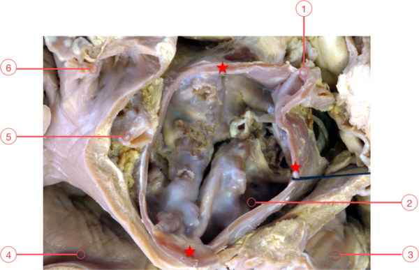
Figure 8. Calcific aortic stenosis
1 - Right coronary office
2 - Non-adjacent sinus
3 - Right atrium
4 - Left atrium
5 - Left coronary artery
6 - Left atrial appendage
This view is looking into the left ventricular outflow from the apex. The aorta to mitral fibrous continuity is thickened and has a redundant, shelf-like appearance secondary to the calcific nature of the aortic valvar leaflets. The leaflets are focally nodular and ulcerated with a slit-like, slightly eccentric opening. The normal semilunar nature of the leaflets and the overall anatomy of the aortic root have been disrupted.
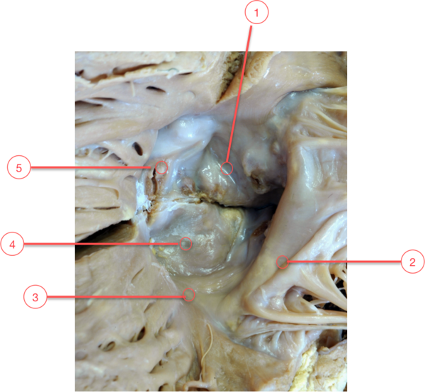
Figure 9. Calcific aortic stenosis
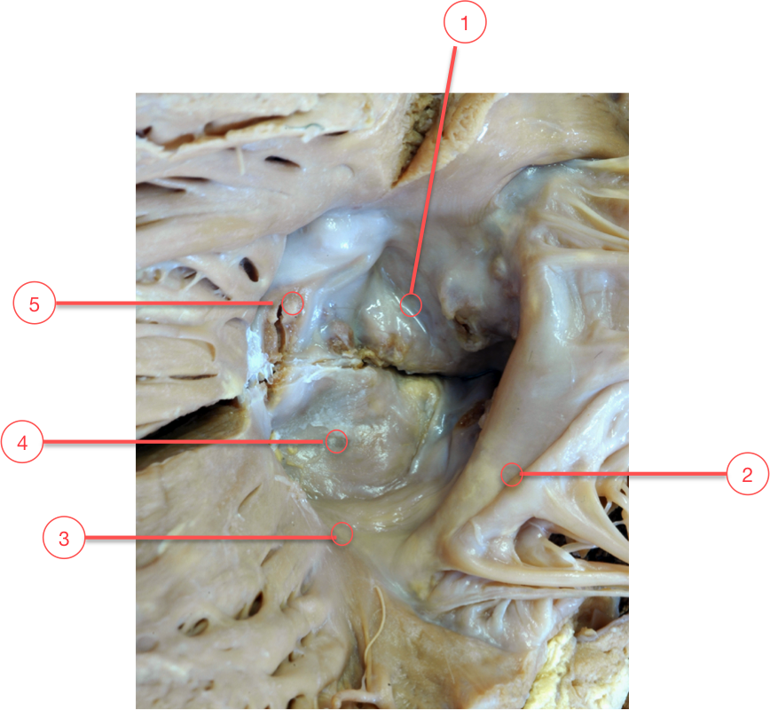
1 - Left coronary leaflet
2 - Mitral valve
3 - Membranous septum
4 - Non-facing leaflet
5 - Right coronary leaflet
Ventricular aspect of aortic and mitral valves
The non-adjacent leaflet and part of the left coronary leaflet are in fibrous continuity with the aortic or anterior leaflet of the mitral valve. At either end, this area of fibrous continuity thickens to form the right and left fibrous trigones. The right fibrous trigone itself is then continuous with the membranous septum, and together these structures form the central fibrous body. When considered together, the fibrous trigones serve to anchor the aorto-mitral valvar unit to the roof of the left ventricle (Figure 10).
Inadvertent low transcatheter heart valve (THV) implantation of a prosthetic valve can, therefore, impinge on the aortic leaflet of the mitral valve, and yield mitral valve dysfunction13.
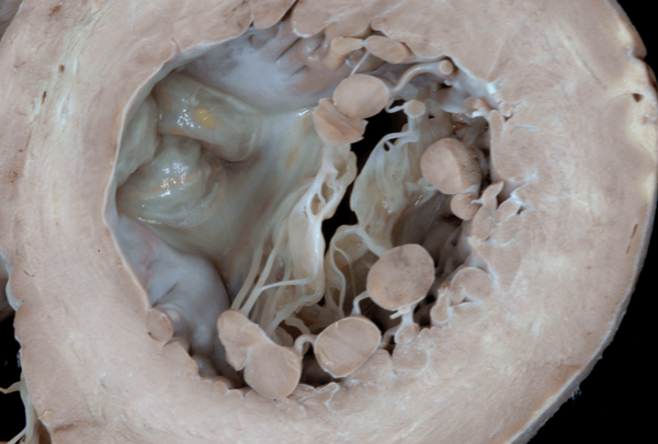
Figure 10. Ventricular aspect of aortic and mitral valves
In this simulated, short-axis, apical, echocardiographic view, the aortic root lies within the central portion of the heart and lifts the mitral valve away from the muscular, interventricular septum. The mitral valve is a bifoliate structure and is supported by paired papillary muscles with no cordal attachments to the septum.
The aorta to mitral fibrous continuity supports approximately one-third of the aortic root with the remaining two-thirds supported by muscle. This area of fibrous continuity is quite strong and is supported by the thickened areas of the left and right fibrous trigones. The right and left fibrous trigones are easily seen at each end of the fibrous continuity with the interleaflet triangle (red lines) extending between the zones of apposition between the left aortic sinus and the non-adjacent aortic sinus.
The non-adjacent sinus is entirely supported by the area of fibrous continuity between the aortic and mitral valves. The right fibrous trigone joins with the membranous septum at the base of the interleaflet triangle between the right aortic sinus and the non-adjacent sinus and forms the central fibrous body.
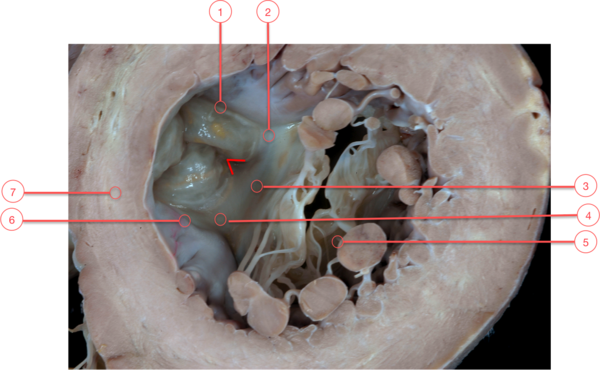
Figure 10. Ventricular aspect of aortic and mitral valves

1 - Aortic valve
2 - Left fibrous trigone
3 - Aorta to mitral continuity
4 - Right fibrous trigone
5 - Mitral valve
6 - Membranous septum
7 - Interventricular septum