Sinotubular junction
Anatomy of the Aortic Valvar Complex
The sinotubular junction is the region of the ascending aorta between the aortic sinuses (of Valsalva) and where the normal tubular configuration of the aorta is attained.
The superior attachments of the aortic valvar leaflets demarcate the level of the sinotubular junction. This junction marks the exit of the aortic root and the beginning of the ascending aorta (Figure 2, Figure 4). Echocardiographic studies have demonstrated that the diameter of the sinotubular junction is significantly larger in patients with aortic stenosis than in normal patients10.

Figure 4. Sinotubular Junction
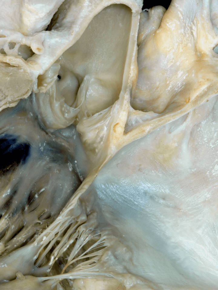
This long-axis, close-up view of the inlet and outlet components of the left ventricle highlights the relationship of the interleaflet triangles to the outside of the heart. The apex of the interleaflet triangles is the most distal extent of where the semilunar hinges of the leaflets join the sinotubular junction (yellow dots).
The interleaflet triangle between the non-adjacent and the right coronary aortic sinus is illustrated with (yellow lines). The (red lines) mark the interleaflet triangle between the non-adjacent and the left coronary aortic sinus, showing nicely how the distal extension of the interleaflet triangles separates the cranial extension of the aortic root from the pericardial cavity. The (white dots) mark the margin of the serous pericardium lining the transverse sinus. The serous pericardium reflects from the epicardial surface of the tubular aorta onto the anterior wall of the atrial chambers and incorporates a small area outside the heart, but within the pericardial cavity. The proximal aspects of two of the interleaflet triangles are in direct contact with the area of the aorta to mitral fibrous continuity and are an integral part of the left and right fibrous trigones. Note the (black dots) which mark the horseshoe-like structure representing the overlapping of the flap valve of the oval fossa with the superior interatrial fold.
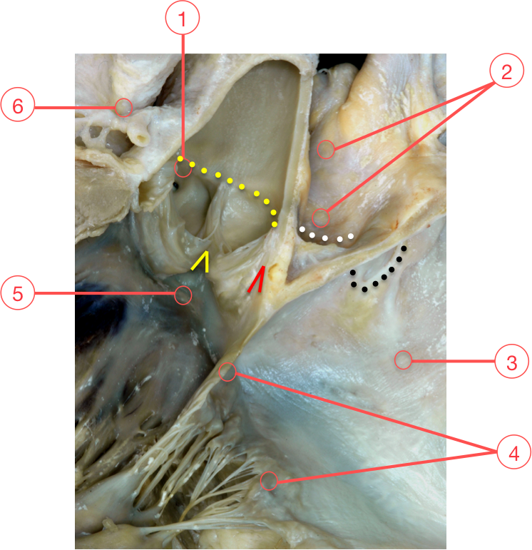
Figure 4. Sinotubular Junction
1 - Right coronary orifice
2 - Transverse sinus
3 - Left atrium
4 - Mitral valve
5 - Membranous septum
6 - Left coronary artery