Relationship to the conduction system
The conducting system of the heart consists of cardiac muscle cells and conducting fibres (not nervous tissue) that are specialised for initiating impulses and conducting them rapidly through the heart. Find out more...
Triangle of Koch
In the right atrium, the atrioventricular node is located within the triangle of Koch. The boundaries of this triangle are the tendon of Todaro, the orifice of the coronary sinus, and the attachment of the septal leaflet of the tricuspid valve (Figure 17). The hinge point of the septal leaflet of the tricuspid valve separates the membranous septum into its atrioventricular and interventricular components. It is the atrioventricular component of this membranous septum that forms the apex of the triangle of Koch, with the atrioventricular node found just inferior to the apex of this triangle.
This heart is viewed from the right side, illustrating the component parts of the triangle of Koch.

Figure 17. Triangle of Koch

Atrioventricular node
The atrioventricular node continues as the bundle of this, which penetrates the atrioventricular component of the membranous septum and runs superficially along the crest of the ventricular septum to give rise to the fascicles of the left bundle branch. The left bundle is closely related to the base of the interleaflet triangle between the right and non-adjacent leaflets of the aortic valve, with the superior part of the bundle intimately related to the right coronary aortic leaflet (Figure 18)23.
Autopsy findings suggest that the average distance between the nadir of the non-adjacent aortic valve leaflet and the left bundle branch is 6.3 ± 2.7 mm. Thus, the conduction axis is closely related to the subaortic apparatus and can be injured during or after balloon valvuloplasty or TAVI.
For more information, consult Conduction disturbances and arrhythmia [link]

Figure 18. Atrioventricular node
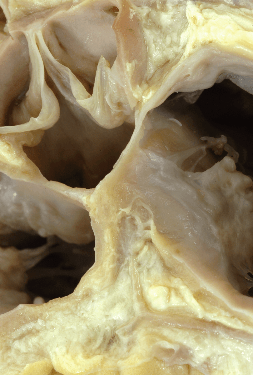
This heart is viewed from the short axis at the base of the heart and tilted a bit to the left. The atrial musculature has been removed along with the non-adjacent sinus of the aortic valve. The right (red star) and left (blue star) facing sinuses remain intact. The area of aortic to mitral fibrous continuity is marked by the red line and the image illustrates how the subaortic outflow tract is lifting the mitral valve away from the interventricular septum.
On the opposite aspect, a portion of the septal leaflet of the tricuspid valve has been removed to view the atrioventricular (yellow dots) and the interventricular (red dots) components of the membranous septum. This demonstrates the relationship between the base of the right interleaflet triangle, the right fibrous trigone and the membranous septum.
The red diamond marks the site of the atrioventricular conduction axis. Note the artery to the atrioventricular node within the fat that interposes between the right and left atrial walls and the crest of the muscular ventricular septum at the base of the inferior interatrial fold.

Figure 18. Atrioventricular node
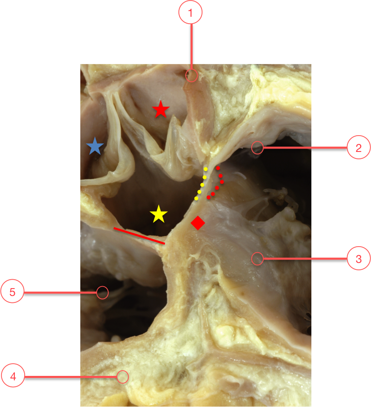
1 - Right coronary orifice
2 - Medical papillary muscle
3 - Tricuspid valve
4 - Artery to the atrioventricular node
5 - Mitral valve