The aortic root
Anatomy of the Aortic Valvar Complex
Understanding the anatomy of the aortic root is particularly relevant in the current era of evolving management strategies for device implantations.
The "aortic root" refers to the aortic outflow tract from its entrance at the left ventricular outlet to its junction with the ascending portion of the aorta7.
It is demarcated inferiorly by a virtual plane created by joining the basal attachment of the aortic valvar leaflets within the left ventricular outflow tract, and superiorly by the distal attachment of the leaflets at the sinotubular junction (Figure 1)8. Within the root as thus defined, it is possible to recognise the sinuses of Valsalva, the sinotubular junction, the fibrous interleaflet triangles, and the semilunar valvar leaflets with their attachments in part to the ventricular and aortic walls, and in part to the aortic or anterior leaflet of the mitral valve (Figure 2). The root lies posterior and rightward relative to the subpulmonary infundibulum, with its circumference bordered anteriorly by the muscular left ventricle, and posteriorly by the orifices of the atrioventricular valves.

Figure 1. The aortic root

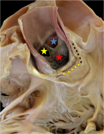
Figure 2. The aortic root
The left atrial and left ventricular walls along with the posterior wall of the aortic root have been removed to show the relationships of the structures surrounding the aortic root.
Anteriorly, the aortic root is bordered by the pulmonary trunk, the two arterial trunks spiralling as they leave the ventricular mass and exit the pericardial cavity. The pulmonary trunk bifurcates into the right and left pulmonary arteries and the arterial ligament extends from the base of the left pulmonary artery to the underside of the aortic arch.
Adjacent to the pulmonary trunk and along its left posterior aspect, the left coronary artery arises from the aortic root and from the left facing or left coronary aortic sinus.
The right facing or right coronary aortic sinus gives rise to the right coronary artery with the right coronary orifice arising just below the sinotubular junction in this specimen. The aortic root extends posterior and to the right of the pulmonary trunk with the most distal extension of the root marked by the sinotubular junction (yellow dots) and giving way to the tubular or ascending aorta.
The ascending aorta then becomes the transverse aortic arch from which the brachiocephalic arteries arise.
The area between the left subclavian artery, the most distal of the brachiocephalic arteries, and the arterial ligament is the aortic isthmus. The aortic isthmus then continues as the descending aorta.
The right pulmonary artery crosses behind the ascending aorta on its way to the root of the right lung. The superior caval vein flanks the ascending aorta on the right.
Within the left atrium, the horseshoe-like structure (red dots) marks the posterior aspect of the flap valve of the oval fossa where it overlaps the superior interatrial fold. The anterior walls of the right and left atrium lie adjacent to the aortic root and the space separating them is intrapericardial, known as the transverse sinus (white dots).
The inlet of the left ventricle is guarded by the mitral valve with the anterior or aortic leaflet separating the inlet from the outlet component. This leaflet (yellow line) is in fibrous continuity with the aortic valve and forms an integral component of the supporting structures for the aortic root.
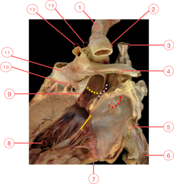
Figure 1. The aortic root
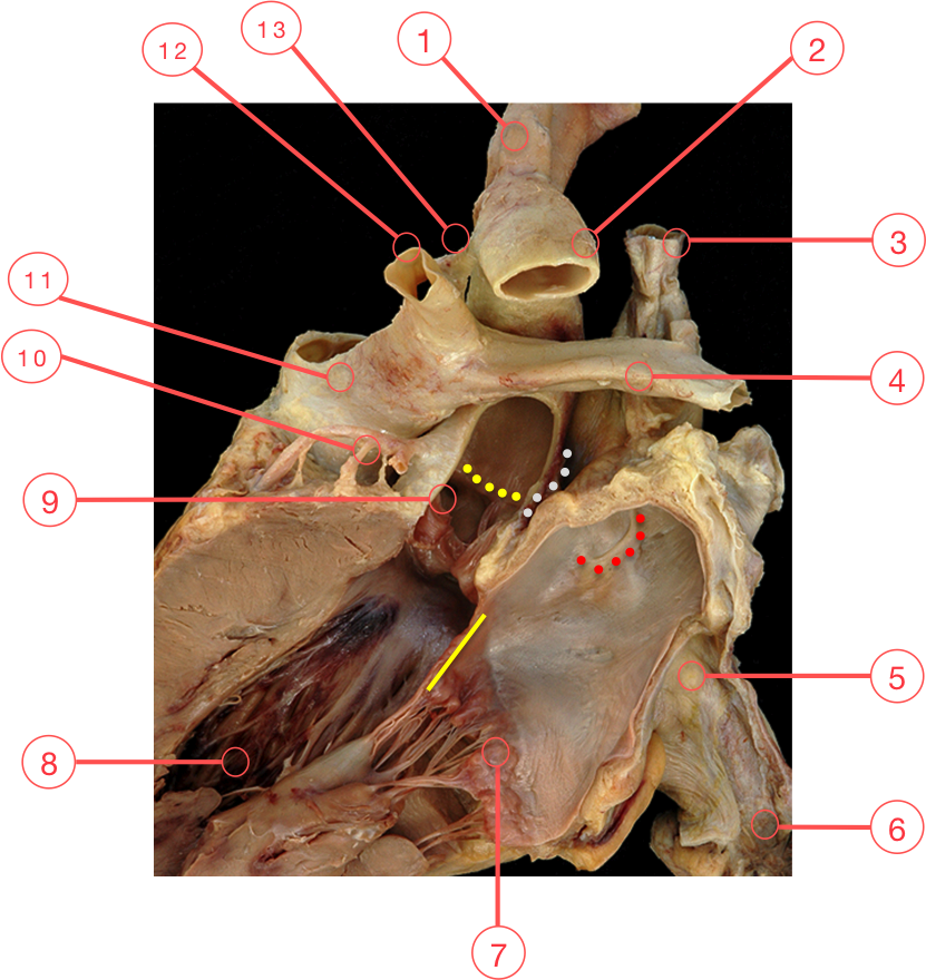
1 - Brachiocephalic arteries
2 - Descending aorta
3 - Superior caval vein
4 - Right pulmonary artery
5 - Left atrium
6 - Inferior caval vein
7 - Mitral valve
8 - Left ventricle
9 - Right coronary orifice
10 - Left coronary artery
11 - Pulmonary trunk
12 - Left pulmonary artery
13 - Arterial ligament
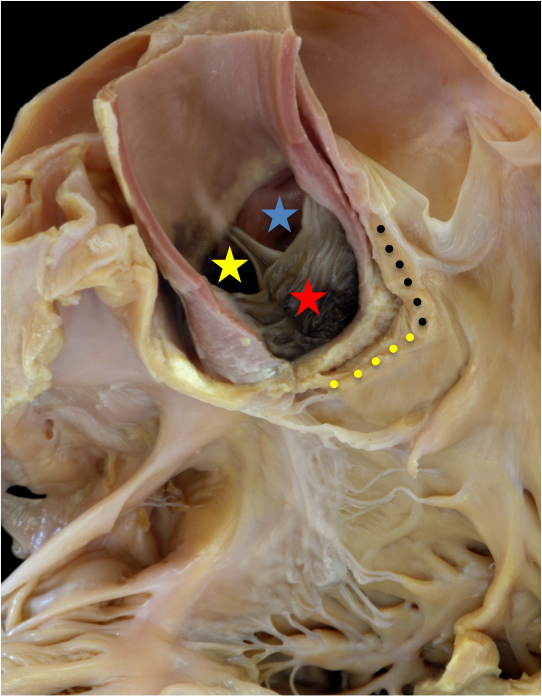
This is a simulated, oblique, subcostal echocardiographic view demonstrating the central position of the aortic root relative to the other cardiac valves.
The aortic valve is to the right and posterior to the pulmonary valve which is supported by a complete, subpulmonary muscular infundibulum (black dots).
Two leaflets of the aortic valve are immediately adjacent to the pulmonary trunk with those aortic sinuses typically giving rise to the coronary arteries.
The red star marks the right-hand facing sinus and gives origin to the right coronary artery, while the left-hand facing sinus (blue star) gives origin to the left main coronary artery. The yellow star marks the non-adjacent aortic sinus which has been referred to in the past as the non-coronary aortic sinus.
Although rare, coronary arteries have been known to arise from this sinus, so the best reference to this sinus is non-adjacent. The leaflets of the aortic valve attach to the aortic wall at the sinotubular junction which is the most distal margin of the aortic root.
On the anterior aspect of the aortic root lies the inner heart curvature which is at the junction of the subpulmonary infundibulum (black dots) and the parietal wall of the right ventricle, this area is sometimes referred to as the ventriculo-infundibular fold (yellow dots).
Note the close proximity of the right atrial musculature to the right of the aortic root.
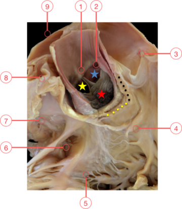
Figure 2. The aortic root
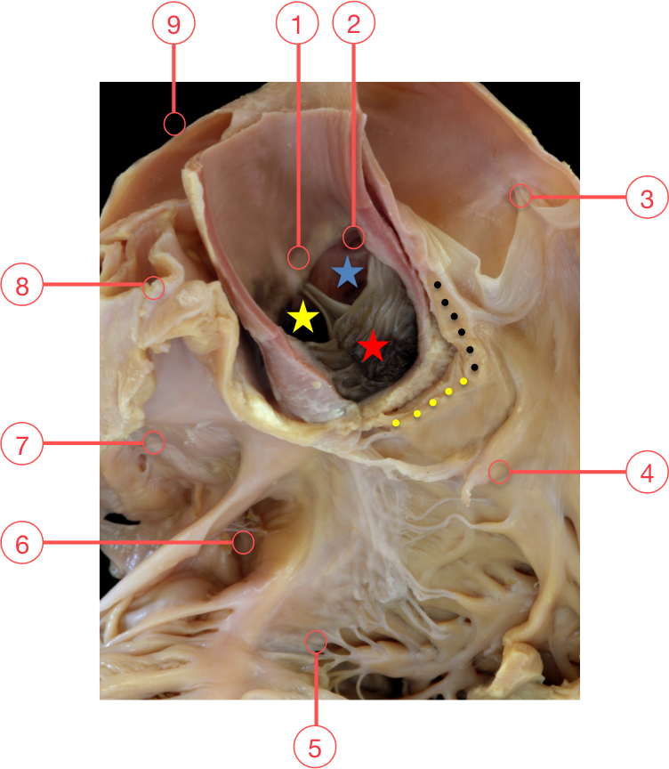
1 - Sinotubular junction
2 - Left coronary orifice
3 - Pulmonary valve
4 - Medical papillary muscle
5 - Tricuspid valve
6 - Coronary sinus
7 - Oval fossa
8 - Superior caval vein
9 - Right pulmonary artery