198 results
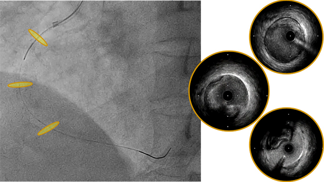
Catheter-based left atrial appendage closure guided by intracardiac echocardiography in a patient with recurrent esophageal variceal bleedings
06 Jul 2026
A 76-year-old patient with permanent atrial fibrillation and recurrent oesophageal variceal bleeding can no longer receive oral anticoagulation, leaving stroke prevention as a major challenge. How would you manage this complex balance between thromboembolic and bleeding risk?
Frequency of the problem:
Expert level:

Author
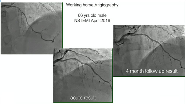
An unconventional pathway to the management of an iatrogenic dissection
23 Jun 2026
Iatrogenic coronary dissection is a rare but potentially life-threatening complication of PCI. Its management can be challenging, particularly when antegrade wiring fails and flow is compromised, requiring escalation to advanced bailout strategies.

Author
DCB: what we know and what we do not know
22 May 2026 – From EuroPCR 2026
This session provides a detailed update on drug-coated balloons (DCB) in interventional cardiology, covering the latest clinical data and trials. It identifies scenarios where DCB use is most effective and offers guidance on optimizing procedural techniques to enhance clinical outcomes.
Multivessel disease or left main disease
22 May 2026 – From EuroPCR 2026
This session explores revascularisation strategies for patients with multivessel or left main coronary artery disease. It emphasizes the role of intravascular physiology and imaging in guiding treatment decisions and highlights procedural factors that contribute to improved long-term clinical outcomes, supported by complex case discussions.

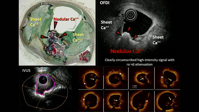
All you need to know - Intracoronary imaging for vulnerable plaque
22 May 2026 – From EuroPCR 2026
This session provides a comprehensive overview of vulnerable plaque pathophysiology and the role of intracoronary imaging in its identification. It assesses treatment options, including the potential role of preventative PCI and intensive medical therapy, to guide clinical decision-making in managing plaque vulnerability.
Spontaneous coronary artery dissection management with intracoronary imaging
22 May 2026 – From EuroPCR 2026
Explore the nuanced management of spontaneous coronary artery dissection (SCAD) with a focus on intracoronary imaging. This session examines diagnostic challenges, the limited role of coronary CT angiography, and strategies for managing occlusive SCAD cases, highlighting the evolving understanding of this complex condition.

Case-based challenges and dilemmas in multivessel PCI
21 May 2026 – From EuroPCR 2026
This session addresses real-world challenges in multivessel PCI for stable and acute coronary syndromes, emphasizing decision-making, procedure planning, and complication management. It highlights the application of imaging, physiology, and contemporary tools such as DCB to optimize outcomes through illustrative cases from Bangladesh, Brunei Darussalam, and the...
Complex multivessel disease: from calcium to physiology - Case-based decisions
21 May 2026 – From EuroPCR 2026
Focus on complex multivessel coronary artery disease management with an emphasis on severely calcified lesions and the integration of intracoronary imaging and physiology. This session prepares clinicians to anticipate and manage complications during PCI, enhancing competence in treating challenging coronary anatomies through case-based decision-making.
Insight from new evidence for complex PCI: value of imaging (CHIP IVUS) and left ventricular support (CHIP BCIS 3)
21 May 2026 – From EuroPCR 2026
This session reviews the impact of recent evidence on complex PCI, highlighting the value of intracoronary imaging (CHIP IVUS trial) and left ventricular assist devices (CHIP BCIS 3 trial). Through case discussions, participants reflect on how these innovations influence daily clinical practice and enhance procedural planning...
CT and intracoronary imaging evaluation of coronary disease
21 May 2026 – From EuroPCR 2026
This session focuses on advanced imaging modalities for evaluating coronary artery disease, including the clinical application of CT angiography and intracoronary imaging techniques like OCT and IVUS. Topics encompass barriers to OCT usage, the impact of plaque composition on PCI outcomes, advances in photon-counting CT technology,...