22 May 2025
PCI for complex calcific coronary artery disease - LIVE Case
Highly calcified lesion preparation guided by OCT before stenting
Summary
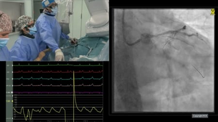
An 87-year-old female patient, at high bleeding risk and with preserved LV function, presented 2 months ago with an inferior STEMI and multivessel disease. She initially underwent RCA PCI followed by staged LCx PCI. She was now referred for PCI of a long, heavily calcified lesion involving the distal left main and mid LAD. Lesion preparation included rotational atherectomy for the mid LAD and IVL for the distal left main/proximal LAD, guided by OCT. Two stents were implanted following adequate lesion preparation.
LIVE Educational Case from Sant'Andrea University Hospital - Rome, Italy
Navigate the video by moving your mouse over the chapter icon in the toolbar

Key moments
- 10:35-13:31 - Imaging analysis
- 19:30-26:05 - Rotablator 1.25 step by step
- 43:00-54:00 - IVL 3.5 step by step
- 32:00-36:38, 40:09-41-09, 1:00:28-1:01:52, 1:15:08-1:20:31 - OCT runs
- 1:23:38-1:27:40 - Procedural analysis
Keywords: Rotablator - IVL - OCT
Learning Objectives
- To discuss about an algorithmic approach to optimise PCI outcome for calcified coronary lesions
- To highlight the pivotal role of intracoronary imaging in procedural planning and device selection during calcified lesions PCI
- To demonstrate the impact of rotational atherectomy and/or intravascular lithotripsy in concentric calcified lesions and calcified nodules


